. 2009 Apr 28;106(17):6950-5.
doi: 10.1073/pnas.0808722106. Epub 2009 Apr 9.

The protein disulfide isomerase AGR2 is essential for production of intestinal mucus

Affiliations

The protein disulfide isomerase AGR2 is essential for production of intestinal mucus

Sung-Woo Park et al. Proc Natl Acad Sci U S A. .

Abstract

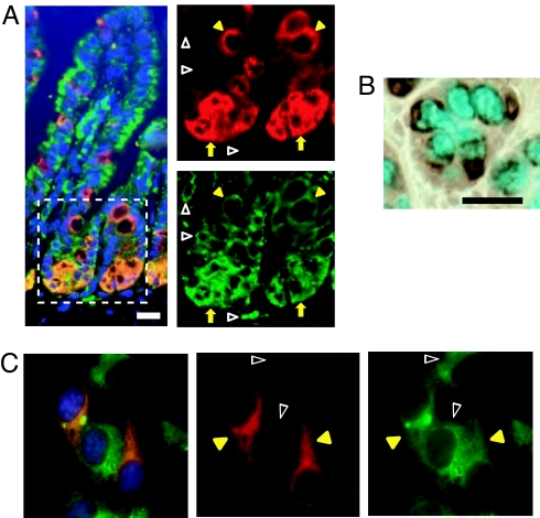
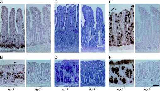
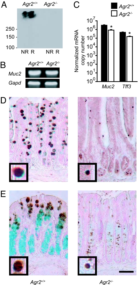
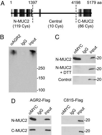
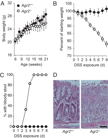
Protein disulfide isomerases (PDIs) aid protein folding and assembly by catalyzing formation and shuffling of cysteine disulfide bonds in the endoplasmic reticulum (ER). Many members of the PDI family are expressed in mammals, but the roles of specific PDIs in vivo are poorly understood. A recent homology-based search for additional PDI family members identified anterior gradient homolog 2 (AGR2), a protein originally presumed to be secreted by intestinal epithelial cells. Here, we show that AGR2 is present within the ER of intestinal secretory epithelial cells and is essential for in vivo production of the intestinal mucin MUC2, a large, cysteine-rich glycoprotein that forms the protective mucus gel lining the intestine. A cysteine residue within the AGR2 thioredoxin-like domain forms mixed disulfide bonds with MUC2, indicating a direct role for AGR2 in mucin processing. Mice lacking AGR2 were viable but were highly susceptible to colitis, indicating a critical role for AGR2 in protection from disease. We conclude that AGR2 is a unique member of the PDI family, with a specialized and nonredundant role in intestinal mucus production.


Conflict of interest statement

The authors declare no conflict of interest.

Figures

Fig. 1.
Fig. 1.
AGR2 is localized to the ER. (A) Mouse intestinal villi were stained with antibodies to AGR2 (red) and the ER proteins GRP78 and GRP94 (green). The outlined area within the composite image at left is magnified in the images on the right to show AGR2 localization in the ER in a subset of cells at the base of the crypt area (yellow arrows) and in goblet cells above the crypts (yellow arrowheads). AGR2 was absent from the ER of other cells (open white arrowheads). (Magnification: 400×.) (B) AGR2 staining (brown) surrounding mucous granules in the colon. Mucus was stained with Alcian blue. (Scale bar: 25 μm.) (C) COS7 cells were transfected with AGR2-FLAG and stained with antibodies to FLAG (red) and the archetypical PDI (P4HB; green). DAPI (blue) was used to visualize nuclei. (Left) A composite image. Yellow arrowheads indicate cells expressing both AGR2-FLAG and P4HB, and open white arrowheads indicate cells expressing P4HB but not AGR2-FLAG. (Magnification: 400×.)
Fig. 2.
Fig. 2.
Agr2−/− mice are devoid of intestinal mucus. (A and B) Immunohistochemical detection of AGR2 protein in small intestine (A) and colon (B) of Agr2+/+ and Agr2−/− mice. (C and D) Periodic acid-Schiff staining of glycoproteins in small intestine (C) and colon (D). (E and F) Immunohistochemical detection of MUC2 protein in small intestine (E) and colon (F). (Scale bar: 50 μm.)
Fig. 3.
Fig. 3.
AGR2 is required for production of MUC2 protein but not for specification of the goblet cell lineage. (A) Immunoblotting for MUC2 protein in nonreduced (NR) and reduced (R) samples from small intestine of Agr2+/+ and Agr2−/− mice. (B) Detection of Muc2 mRNA in small intestine by reverse transcription followed by PCR. Gapd mRNA was used as a control. (C) Expression of Muc2 and Tff3 mRNAs in small intestine from Agr2+/+ and Agr2−/− mice was measured by quantitative RT-PCR. Results are means ± SEM for 4 mice per group. *, P < 0.001 compared to Agr2+/+ mice. (D and E) TFF3 protein (brown) in the small intestine (D) and colon (E) was identified by immunohistochemistry. Mucus was stained with Alcian blue. (Scale bars: 50 μm.) Insets at the lower left corner of each photomicrograph (3× higher magnification) show typical TFF3-stained granules.
Fig. 4.
Fig. 4.
AGR2 forms mixed disulfide bonds with MUC2. (A) Schematic representation of MUC2, showing the distribution of cysteine residues (vertical lines) in the amino-terminal and carboxyl-terminal, cysteine-rich regions and the central region, which contains relatively few cysteine residues and is composed mainly of a large number of tandem repeats of a heavily glycosylated, threonine-rich sequence. (B) AGR2 was immunoprecipitated from LS174T cells, and associated MUC2 was detected by immunoblotting. IgG from nonimmune serum was used as a control. A total of 4% of the volume of lysate used for the immunoprecipitation was run on the gel for comparison (input). (C) HEK293T cells were cotransfected with an MYC-tagged amino-terminal fragment of MUC2 (N-MUC2) and AGR2-FLAG. N-MUC2-containing complexes were immunoprecipitated with anti-MYC, and associated AGR2 was detected by immunoblotting with anti-FLAG antibody. AGR2 did not coimmunoprecipitate with N-MUC2 if cells were treated with the reducing agent DTT before lysis. An MYC-tagged construct lacking the N-MUC2 fragment (EGFP-MYC) was used as a control (Bottom). (D) FLAG-tagged AGR2 coimmunoprecipitated with MYC-tagged amino-terminal and carboxyl-terminal fragments of MUC2, but a FLAG-tagged AGR2 mutant lacking the cysteine residue (C81S) did not.
Fig. 5.
Fig. 5.
Agr2−/− mice are highly susceptible to DSS-induced colitis. (A) Mean body weights of mice with no DSS exposure (6 mice per group; P > 0.2 for Agr2−/− compared with Agr2+/+ mice at all ages). Error bars represent SEM. (B) Weight change during DSS administration in Agr2−/ − mice (6 mice per group; P < 10−5 at 8 days). (C) Appearance of blood in the stool during DSS administration. Agr2−/ − mice were more likely than control mice to develop bloody stools during low-dose DSS treatment (P = 1 × 10⁻⁵ by Fisher's exact test). (D) Hematoxylin and eosin staining of colon from DSS-exposed mice. (Scale bar: 50 μm.)

References

    1. van Anken E, Braakman I. Versatility of the endoplasmic reticulum protein folding factory. Crit Rev Biochem Mol Biol. 2005;40:191–228. - PubMed
    1. Ellgaard L, Ruddock LW. The human protein disulphide isomerase family: Substrate interactions and functional properties. EMBO Rep. 2005;6:28–32. - PMC - PubMed
    1. Persson S, et al. Diversity of the protein disulfide isomerase family: Identification of breast tumor induced Hag2 and Hag3 as novel members of the protein family. Mol Phylogenet Evol. 2005;36:734–740. - PubMed
    1. Anelli T, et al. ERp44, a novel endoplasmic reticulum folding assistant of the thioredoxin family. EMBO J. 2002;21:835–844. - PMC - PubMed
    1. Anelli T, et al. Thiol-mediated protein retention in the endoplasmic reticulum: The role of ERp44. EMBO J. 2003;22:5015–5022. - PMC - PubMed

Publication types

MeSH terms

Associated data