. 2008 Oct 3;135(1):37-48.
doi: 10.1016/j.cell.2008.09.016.

Transcription factor E2-2 is an essential and specific regulator of plasmacytoid dendritic cell development

Affiliations

Transcription factor E2-2 is an essential and specific regulator of plasmacytoid dendritic cell development

Babacar Cisse et al. Cell. .

Abstract

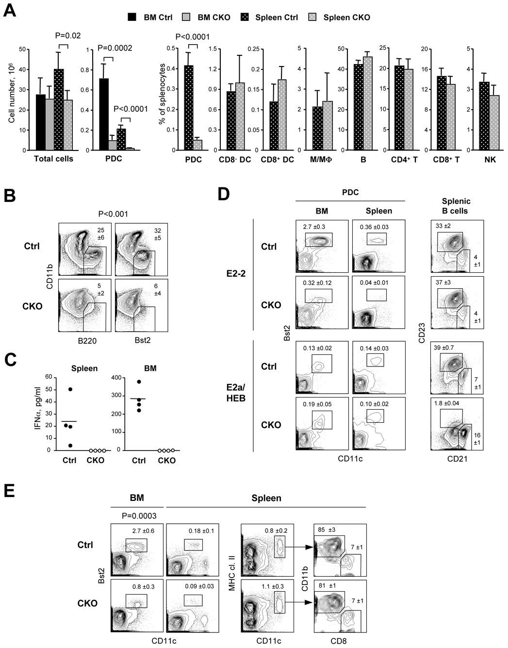
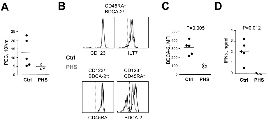
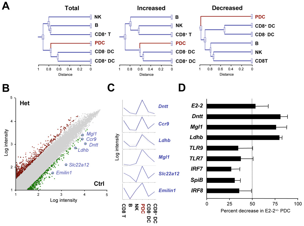
Plasmacytoid dendritic cells (PDCs) represent a unique immune cell type specialized in type I interferon (IFN) secretion in response to viral nucleic acids. The molecular control of PDC lineage specification has been poorly understood. We report that basic helix-loop-helix transcription factor (E protein) E2-2/Tcf4 is preferentially expressed in murine and human PDCs. Constitutive or inducible deletion of murine E2-2 blocked the development of PDCs but not of other lineages and abolished IFN response to unmethylated DNA. Moreover, E2-2 haploinsufficiency in mice and in human Pitt-Hopkins syndrome patients was associated with aberrant expression profile and impaired IFN response of the PDC. E2-2 directly activated multiple PDC-enriched genes, including transcription factors involved in PDC development (SpiB, Irf8) and function (Irf7). These results identify E2-2 as a specific transcriptional regulator of the PDC lineage in mice and humans and reveal a key function of E proteins in the innate immune system.


Figures

Figure 1
Figure 1. E2-2 is preferentially expressed in the PDC
(A) Expression of E protein genes in sorted splenic cell populations from wild-type mice, including PDC (CD11clow Bst2+), B cells (CD11c B220+), T cells (TCRβ+), monocytes/macrophages (M, CD11c CD11b+ side scatterlow) and cDC (CD11chigh CD11b+). Data represent normalized expression levels relative to the PDC sample, as determined by qRT-PCR (mean ±S.D. of triplicate reactions). The expression of the PDC-enriched transcription factor SpiB is shown as a control. (B) Expression of E protein genes in sorted human peripheral blood cell populations, determined by qRT-PCR as above. Cells include PDC (BDCA-4+), B cells (CD19+), T cells (CD3+), monocytes (Mo, CD14+) and total mononuclear cells (MNC). (C) The expression of E proteins and Id2 in PDC. Shown is RT-PCR analysis of cell populations sorted as described in panel A. (D) The expression of E2-2 in PDC undergoing differentiation into cDC. BM culture-derived PDC were purified, incubated with type B CpG for 48 hr (+), and analyzed by qRT-PCR for the expression of E2-2 and SpiB relative to the untreated PDC (−). The expression of a cDC-specific pI transcript of class II transactivator (CIITA pI) is shown as a control for DC differentiation. (E) The expression of E proteins in the human PDC cell line. Shown is Western blot analysis of the indicated proteins in the PDC line CAL-1 and in T cell lymphoma MOLT-4. (F) The analysis of E box-binding nuclear proteins in the PDC. Shown is EMSA using radiolabeled E box-containing probe and nuclear extracts from CAL-1 or MOLT-4 cells. The binding reactions contained no additives (−), control (Ctrl) or E protein-specific antibodies, unlabeled probe as a competitor (Comp), or unlabeled probe with a mutated E box (Comp*). No supershift was observed with anti-HEB antibody in CAL-1 (not shown).
Figure 2
Figure 2. E2-2 deficiency blocks PDC development
(A) The analysis of hematopoietic chimeras reconstituted with E2-2+/+ control (Ctrl) or E2-2−/− knockout (KO) fetal liver cells. Shown is the fraction of donor-derived (CD45.2+) cells in the total BM and spleen, and the fractions of indicated cell types among the donor-derived CD45.2+ cells. Cells included PDC (CD11clow Bst2+), B cells (CD11c B220+), monocytes/macrophages (CD11c CD11b+ side scatterlow), granulocytes (CD11b+ side scatterhigh) and cDC (CD11chigh MHC cl. II+ CD11b+ or CD11b). Data represent mean percentage ±S.D. of 3 control and 5 KO chimeras. (B) Representative staining profiles of donor-derived (CD45.2+) spleen and BM cells, with the PDC population indicated. (C) PDC development in Flt3L-supplemented BM cultures from control or E2-2 KO-reconstituted chimeric mice. Shown are representative staining profiles of donor-derived CD11c+ cells at the indicated days of culture. (D) The analysis of PDC population in the BM of control or E2-2 KO-reconstituted chimeric mice. Left panel shows representative staining profiles of CD45.2+ total BM cells, with the PDC-containing CD11c+ CD11bpopulation indicated. Right panel shows fluorescence histograms of CD45.2+ CD11c+ CD11b cells stained for the indicated surface markers; dotted line indicates positive staining threshold. (E) A model of sequential PDC development in the mouse BM, showing pro-DC (open circle), immature PDC (light grey), and mature lineage marker-positive PDC (dark grey). Immature PDC were low/negative for M-CSFR and SiglecH (data not shown). The developmental block caused by E2-2 deletion is indicated.
Figure 3
Figure 3. Conditional inactivation of E2-2 impairs PDC development
(A) The analysis of R26-CreER+ control (Ctrl, E2-2wt/wt) and conditional knockout (CKO, E2-2flox/flox) mice 10 days after the induction of Cre recombination by tamoxifen. Shown are absolute numbers of total cells and of the PDC in the spleen and BM, and fractions of indicated cell types in the spleen (mean ±S.D. of 4 animals per genotype). Cells included PDC (CD11clow Bst2+), B cells (CD11c B220+), monocytes/macrophages (M/MΦ, CD11c CD11b+ side scatterlow), cDC (CD11chigh MHC cl. II+, CD8 or CD8+), T cells (TCRβ+, CD4+ or CD8+) and NK cells (DX5+). (B) PDC development in Flt3L-supplemented BM cultures from induced control and CKO mice. Shown are representative staining profiles of CD11c+ cells from day 9 BM cultures; CD11b B220+ or Bst2+ PDC are indicated (mean percentage ±S.D. of cultures from 4 individual mice). (C) CpG-induced secretion of IFNα by total splenocytes and BM cells from induced control and CKO mice. Cells were incubated with type A CpG for 48 hr, and IFNα concentration in the supernatant was determined by ELISA. Symbols represent cell cultures from individual mice. (D) Tamoxifen-induced deletion of E proteins in adult animals. R26-CreER+ mice conditional for E2-2 (E2-2flox/flox) or E2a/HEB (E2aflox/flox HEBflox/flox) were analyzed 10 days after the induction. Wild-type C57BL/6 mice were used as controls for E2a/HEB CKO. Shown are gated B220+ BM or spleen cells stained for PDC (CD11clow Bst2+) or gated B220+ AA4.1 splenocytes stained for follicular (CD23highCD21low) or marginal zone (CD23lowCD21high) B cells. The fractions of cell populations among total BM or spleen cells are indicated (mean ±S.D. of 2–3 animals per genotype). (E) The analysis of CD11c-Cre+ control (E2-2 wt/wt) and CKO (E2-2flox/flox) animals. Shown are representative staining profiles of total BM and spleen cells from control and CKO mice, with the percentage of PDC, cDC and cDC subsets indicated (mean ±S.D. of 5 animals per genotype).
Figure 4
Figure 4. Impaired PDC development and function in E2-2 heterozygous mice
(A) Representative staining profiles of PDC from the BM and spleen of E2-2+/− mice (Het) and wild-type control (Ctrl) littermates. (B) Absolute numbers of PDC (CD11clow Bst2+) in E2-2+/− and control mice (mean ±S.D. of 8–11 mice per genotype). The numbers of splenic B cells (B220+) and cDC (CD11chigh MHC cl. II+) are also shown. (C) PDC development in Flt3L-supplemented BM cultures from E2-2+/− and control mice. Shown are representative staining profiles of gated CD11c+ cells from day 10 BM cultures; the PDC fraction is indicated (mean percentage ±S.D. of cultures from 4 individual mice). (D) Induction of IFNβ gene expression in vivo. E2-2+/− mice or E2-2+/+ littermate controls carrying one copy of IFNβ-EYFP knock-in allele were injected with CpG/lipid complex, and EYFP expression was measured 6 hr later. Shown are EYFP fluorescence profiles of splenic PDC (B220+ Bst2+), with the percentage and mean fluorescence intensity (MFI) of EYFP+ cells indicated. Representative of four independent experiments (5–7 animals per genotype). (E) Systemic IFN response after challenge with TLR ligands in vivo. E2-2+/− or control littermates were injected with TLR9 ligand CpG/lipid or with TLR3 ligand pI:C, and serum IFNα was measured 6 or 12 hr later, respectively. E2-2+/− mice similarly failed to mount IFN response to CpG after 12 hr (not shown). Symbols represent IFNα concentrations in individual animals. The relatively low levels of CpG-induced IFNα are likely due to the early time point analyzed. (F) T cell-dependent antibody responses in E2-2+/− and control mice. The mice were immunized with nitrophenyl (NP)-keyhole limpet hemocyanin (KLH) conjugate (50 µg in alum adjuvant), and serum anti-NP antibody titers were measured by ELISA at the indicated time points. Symbols represent individual control (open squares) or E2-2+/− (black triangles) immunized mice; asterisks indicate antibody levels below the detection limit. Similar results were obtained with IgG2a isotype (not shown).
Figure 5
Figure 5. Impaired PDC phenotype and function in the human PHS patients
Total peripheral blood cells from three PHS patients and five healthy controls were analyzed. (A) Absolute number of PDC (CD45RA+ CD123+ BDCA-2+) in the peripheral blood as determined by flow cytometry. Symbols represent individual patients (PHS) and control subjects (Ctrl). (B) Cell surface marker expression by PDC. Total blood cells were simultaneously stained for the indicated PDC markers; shown are fluorescence histograms of the PDC population (gated as indicated) from a representative control and a PHS patient. (C) The expression level of BDCA-2/CD303 on PDC, shown as mean fluorescent intensity (MFI) of the BDCA-2+ population. (D) The secretion of IFNα in response to CpG in vitro. Total peripheral blood was incubated with type A CpG for 24 hr, and IFNα concentration in the supernatant was determined by ELISA.
Figure 6
Figure 6. E2-2 regulates the expression of PDC-enriched genes
Sorted PDC from E2-2+/− mice and wild-type littermate controls were analyzed by microarray expression profiling, and the results were compared to the expression database of normal immune cell populations (GEO dataset GSE9810). (A) Clustering of cell populations by total probe set or by the probe sets increased or decreased in E2-2+/− PDC. (B) Pairwise comparison of E2-2+/− (Het) and control (Ctrl) PDC expression profiles. The scatter plot represents normalized log intensities of individual probes, with the probes increased or decreased >3-fold in E2-2+/− PDC indicated in red and green, respectively. The probes prominently decreased in E2-2+/− PDC are highlighted in blue. (C) Expression profiles of the decreased probes from panel B in normal immune cell populations. (D) Gene expression in sorted PDC as determined by qRT-PCR. Data represent percent decrease of the indicated genes in E2-2+/− compared to control wild-type PDC (mean ±S.D. of values from 3–4 independently sorted and analyzed PDC samples). The expected 50% decrease of E2-2 expression is indicated by the dotted line.
Figure 7
Figure 7. E2-2 directly activates PDC-enriched genes
(A) The expression of PDC-enriched transcription factors following E2-2 deletion in mature PDC. Purified BM culture-derived PDC from naïve R26-CreER+ control (E2-2wt/wt) or CKO (E2-2flox/flox) mice were incubated with tamoxifen in vitro, and the expression of indicated transcripts was monitored by qRT-PCR. No differences in cell viability have been observed in the cultures (not shown). The levels of all genes at 1 day were similar to those on day 0, and were taken as 100%. Data represent mean normalized values ±S.D. of triplicate PCR reactions. (B) The binding of E2-2 to the regulatory regions of PDC-expressed genes. Chromatin immunoprecipitation (ChIP) was performed in human PDC line CAL-1 using anti-E2-2 or control antibodies, and the isolated chromatin was analysed by qPCR. The data represent normalized fold enrichment of the indicated genes in anti-E2-2 compared to control antibody ChIP (mean ±S.D. of triplicate reactions). An irrelevant gene (PROM1) and an irrelevant internal fragment of CLEC4C gene (CLEC4C*) were used as negative controls. (C) The map of the human SPIB 5’ region and first intron, with the percent identity plot comparison to other genomes generated by ECR software (http://ecrbrowser.dcode.org). The conserved region in the first intron amplified by ChIP is highlighted with a red oval, and partial alignment of the human (top) and mouse (bottom) sequences is shown to the right. Conserved E boxes are indicated in red.

References

    1. Amiel J, Rio M, de Pontual L, Redon R, Malan V, Boddaert N, Plouin P, Carter NP, Lyonnet S, Munnich A, et al. Mutations in TCF4, encoding a class I basic helix-loop-helix transcription factor, are responsible for Pitt-Hopkins syndrome, a severe epileptic encephalopathy associated with autonomic dysfunction. Am J Hum Genet. 2007;80:988–993. - PMC - PubMed
    1. Asselin-Paturel C, Trinchieri G. Production of type I interferons: plasmacytoid dendritic cells and beyond. J Exp Med. 2005;202:461–465. - PMC - PubMed
    1. Bain G, Gruenwald S, Murre C. E2A and E2-2 are subunits of B-cell-specific E2-box DNA-binding proteins. Mol Cell Biol. 1993;13:3522–3529. - PMC - PubMed
    1. Banchereau J, Pascual V. Type I interferon in systemic lupus erythematosus and other autoimmune diseases. Immunity. 2006;25:383–392. - PubMed
    1. Barchet W, Cella M, Colonna M. Plasmacytoid dendritic cells--virus experts of innate immunity. Semin Immunol. 2005;17:253–261. - PubMed

Publication types

MeSH terms

Associated data