. 2024 Jul 15;22(1):363.
doi: 10.1186/s12964-024-01740-5.

Endometrial senescence is mediated by interleukin 17 receptor B signaling

Affiliations

Endometrial senescence is mediated by interleukin 17 receptor B signaling

Keiko Kawamura et al. Cell Commun Signal. .

Erratum in

Abstract

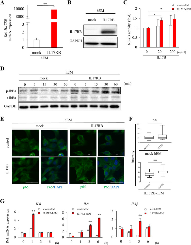
Background: We previously identified Il17RB, a member of the IL17 superfamily, as a candidate marker gene for endometrial aging. While IL17RB has been linked to inflammation and malignancies in several organ systems, its function in the endometrium has not been investigated and is thus poorly understood. In the present study, we performed a functional analysis of this receptor with the aim of determining the effects of its age-associated overexpression on the uterine environment.

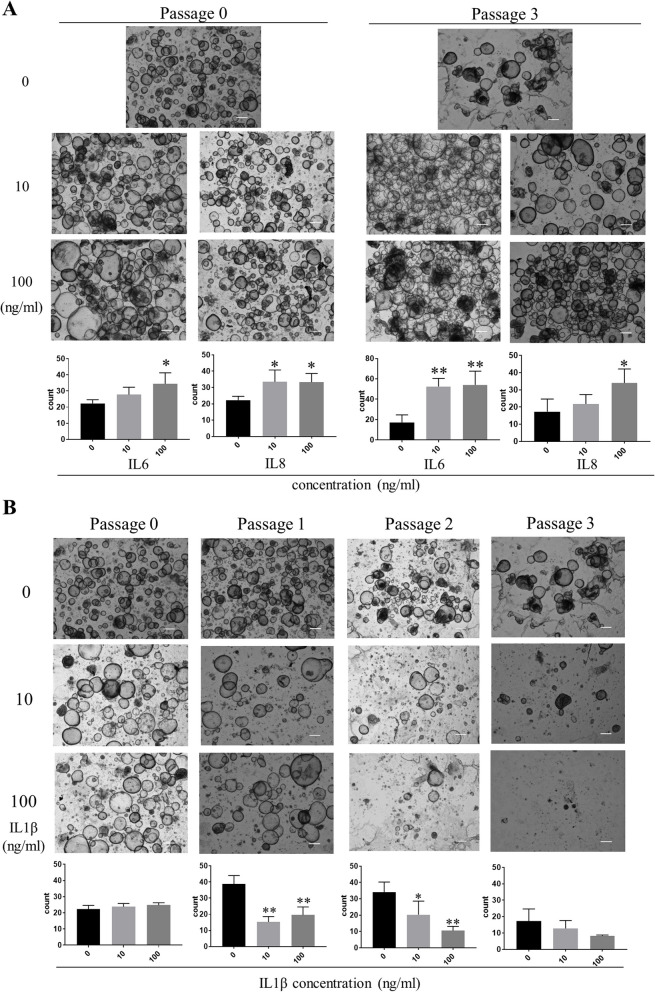
Methods: We analyzed IL17RB-related signaling pathways and downstream gene expression in an immortalized human endometrial glandular epithelial cell line ("hEM") forced to express the receptor via lentiviral transduction ("IL17RB-hEM"). We also prepared endometrial organoids from human endometrial tissue sourced from hysterectomy patients ("patient-derived EOs") and exposed them to cytokines that are upregulated by IL17RB expression to investigate changes in organoid-forming capacity and senescence markers. We analyzed RNA-seq data (GEO accession number GSE132886) from our previous study to identify the signaling pathways associated with altered IL17RB expression. We also analyzed the effects of the JNK pathway on organoid-forming capacity.

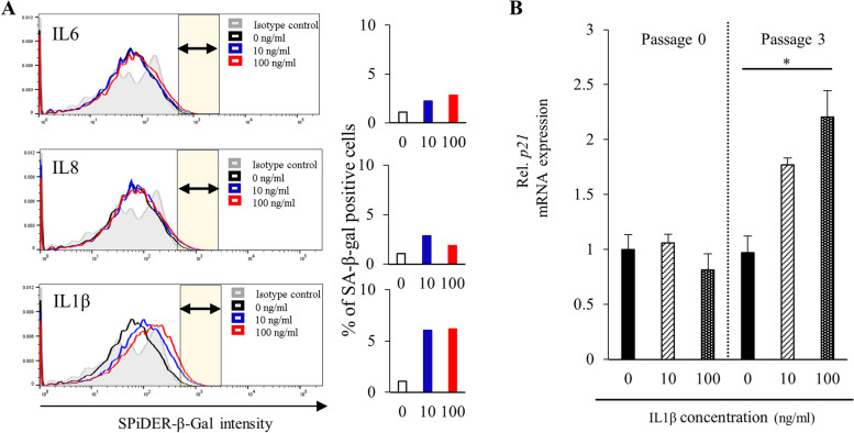
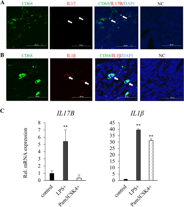
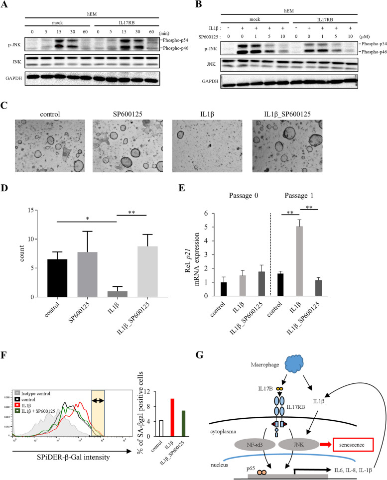
Results: Stimulation with interleukin 17B enhanced the NF-κB pathway in IL17RB-hEM, resulting in significantly elevated expression of the genes encoding the senescence associated secretory phenotype (SASP) factors IL6, IL8, and IL1β. Of these cytokines, IL1β inhibited endometrial organoid growth. Bioinformatics analysis showed that the JNK signaling pathway was associated with age-related variation in IL17RB expression. When IL17RB-positive cells were cultured in the presence of IL17B, their organoid-forming capacity was slightly but non-significantly lower than in unexposed IL17RB-positive cells, but when IL17B was paired with a JNK inhibitor (SP600125), it was restored to control levels. Further, IL1β exposure significantly reduced organoid-forming capacity and increased p21 expression in endometrial organoids relative to non-exposure (control), but when IL1β was paired with SP600125, both indicators were restored to levels comparable to the control condition.

Conclusions: We have revealed an association between IL17RB, whose expression increases in the endometrial glandular epithelium with advancing age, and cellular senescence. Using human endometrial organoids as in vitro model, we found that IL1β inhibits cell proliferation and leads to endometrial senescence via the JNK pathway.

Keywords: Aging; Endometrial senescence; IL17B; IL17RB; IL1β; JNK; Macrophage; NF-κB; Organoid; SASP.


Conflict of interest statement

The authors declare no competing interests.

Figures

Fig. 1
Fig. 1
IL17B/IL17RB signaling pathway analysis (hEM). Experiments compared immortalized human endometrial glandular cells forced to express IL17RB via a lentiviral vector (“IL17RB”) with mock-transfected hEM (“mock”). (A) IL17RB mRNA expression as quantified via RT-qPCR relative to an endogenous control (HPRT1). Error bars denote standard deviation (n = 3). **P < 0.01. (B) IL17RB protein expression as evaluated via Western blotting. GAPDH was used as the loading control. (C) NF-κB activity 6 h after IL17B stimulation as measured via dual luciferase assay. Error bars denote standard deviation (n = 3). *P < 0.05. (D) IκBα phosphorylation after IL17B stimulation (100 ng/ml) as evaluated via Western blotting. p-IκBα: phosphorylated IκBα, t-IκBα: total IκBα, GAPDH: loading control. (E) Immunofluorescent staining of p65 nuclear translocation 16 h after IL17B stimulation (100 ng/ml). Scale bars: 50 μm. (F) Nuclear intensity in p65-immunostained images as measured using ImageJ. Box plots representing the mean intensity per pixel within the nucleus of a single cell are shown. ** P < 0.01, n.s., not significant. (G) IL6, IL8, and IL1β mRNA expression. Expression was measured via RT-qPCR at 0 (baseline), 1, 3, and 6 h after IL17B stimulation (100 ng/ml). Endogenous control: HPRT1. Error bars denote standard deviation (n = 3). **P < 0.01
Fig. 2
Fig. 2
Endometrial response to cytokines downstream of IL17RB (patient-derived EOs). This series of experiments used endometrial organoids grown from tissue specimens from hysterectomy patients. (A) Endometrial organoids (EM_2) cultured in ExM containing IL6 (10, 100 ng/ml) or IL8 (10, 100 ng/ml). Passage 0 and 3 images were taken on day 16 and 17 of culture, respectively. Bar graphs show the numbers of organoids (diameter ≥ 20 μm) counted in each passage and exposure condition (IL6: 0, 10, 100 ng/ml; IL8: 0, 10, 100 ng/ml). Error bars denote standard deviation (n = 4 independent locations). Scale bars: 300 μm. (B) Endometrial organoids (EM_2) cultured in ExM containing IL1β (0, 10, 100 ng/ml). Passage 0, 1, 2, and 3 images were taken on day 16, 10, 11, and 17 of culture, respectively. Bar graphs show organoid count (diameter ≥ 20 μm) by passage and exposure condition (IL1β: 0, 10, 100 ng/ml). Error bars denote standard deviation (n = 4 independent locations). Scale bars: 300 μm
Fig. 3
Fig. 3
A SPiDER-β-Gal flow cytometry of endometrial organoids (EM_2, passage 3). Horizontal axis: SPiDER-β-Gal intensity; vertical axis: unit area. Bar graphs show the percentage of the area under each curve that is brighter than in the negative control (yellow area: SA-β-gal-positive cells). B p21 mRNA expression in patient-derived endometrial organoids (EM_2; passages 0, 3) cultured in ExM containing IL1β (0, 10, 100 ng/ml). Expression was quantified via RT-qPCR relative to an endogenous control (18 S rRNA). Error bars denote standard deviation (n = 3). * P < 0.05, **P < 0.01
Fig. 4
Fig. 4
A Fluorescent double immunostaining (CD68 + IL17B) of sections of healthy endometrial tissue from a female patient in her forties (EM_7). NC: Negative Control. Scale bars: 50 μm. B Fluorescent double immunostaining (CD68 + IL1β) of sections of healthy endometrial tissue from a female patient in her forties (EM_8). NC: Negative Control. Scale bars: 50 μm. (C) IL17B and IL1β mRNA expression measured via RT-qPCR in macrophages derived from peripheral blood monocytes activated with LPS (1000 ng/ml) and Pam3CSK4 (1000 ng/ml). Endogenous control: HPRT1. Error bars denote standard deviation (n = 3). ** P < 0.01
Fig. 5
Fig. 5
A Four Gene Ontology terms among 425 genes having significant associations (P < 0.001) with age-related differences in IL17RB expression in the murine uterus according to Pearson’s correlation analysis (data source: Kawamura et al. 2020). B Western blot time course (60 min) of JNK phosphorylation in IL17RB-hEM cells serum-starved for 16 h and then stimulated with IL17B (100 ng/ml). p-JNK: phosphorylated JNK; t-JNK: total JNK; GAPDH: loading control. C Western blot time course (5 min) of JNK phosphorylation in IL17RB-hEM cells serum-starved for 16 h and then stimulated with IL17B (100 ng/ml) plus SP600125 (0, 1, 5, 10 µM). p-JNK: phosphorylated JNK; t-JNK: total JNK; GAPDH: loading control. DH Phenotypic differences associated with IL17RB expression in human endometrial epithelial cells. D Flow cytometry was used to sort and separate samples of endometrial cells (EM_9) by identifying a region with the classic signature of endometrial glandular cells (CD9+/CD10) and subdividing it based on IL17RB expression. E Endometrial organoids on day 15 of culture. Scale bars: 300 μm. F Organoid count (diameter ≥ 20 μm) on day 15 of culture. Error bars denote standard deviation (n = 8 independent locations). ** P < 0.01. G Endometrial organoid diameter on day 15 of culture. * P < 0.05. H Organoid count (diameter ≥ 20 μm) in IL17RB(+) and IL17RB(−) cells cultured in ExM in the presence of SP600125 (5 µM), IL17B (100 ng/ml), or both. Error bars denote standard deviation (n = 8 independent locations). * P < 0.05
Fig. 6
Fig. 6
A Western blot time course (60 min) of JNK phosphorylation in IL17RB-hEM and mock cells serum-starved for 16 h and then stimulated with IL1β (100 ng/ml). p-JNK: phosphorylated JNK; t-JNK: total JNK; GAPDH: loading control. B Western blot time course (15 min) of JNK phosphorylation in IL17RB-hEM and mock cells serum-starved for 16 h and then stimulated with IL1β (100 ng/ml) plus SP600125 (0, 1, 5, 10 µM). p-JNK: phosphorylated JNK; t-JNK: total JNK; GAPDH: loading control. ce Endometrial organoids (EM_10) cultured in ExM containing SP600125 (5 µM), IL1β (100 ng/ml), or both. C Passage 1 images. Scale bars: 300 μm. D Organoid count (diameter ≥ 20 μm; passage 1). Error bars denote standard deviation (n = 4 independent locations). E p21 mRNA expression (passages 0, 1). Expression was quantified via RT-qPCR relative to an endogenous control (18 S rRNA). Error bars denote standard deviation (n = 3). * P < 0.05, ** P < 0.01. F SPiDER-β-Gal flow cytometry of endometrial organoids (EM_6, passage 2). Horizontal axis: SPiDER-β-Gal intensity; vertical axis: unit area. Bar graphs show the percentage of the area under each curve that is brighter than in the negative control (yellow area: SA-β-gal-positive cells). G Schematic of the endometrial senescence mechanism related to IL17RB

References

    1. Henry L. Some data on natural fertility. Eugen Q. 1961;8:81–91. 10.1080/19485565.1961.9987465 - DOI - PubMed
    1. Katagiri Y, Jwa SC, Kuwahara A, Iwasa T, Ono M, Kato K, Kishi H, Kuwabara Y, Harada M, Hamatani T, Osuga Y. Assisted reproductive technology in Japan: a summary report for 2019 by the ethics committee of the Japan society of obstetrics and gynecology. Reprod Med Biol. 2022;21:e12434. 10.1002/rmb2.12434 - DOI - PMC - PubMed
    1. Franasiak JM, Forman EJ, Hong KH, Werner MD, Upham KM, Treff NR, Scott RT Jr. The nature of aneuploidy with increasing age of the female partner: a review of 15,169 consecutive trophectoderm biopsies evaluated with comprehensive chromosomal screening. Fertil Steril. 2014;101:656–e663651. 10.1016/j.fertnstert.2013.11.004 - DOI - PubMed
    1. Rubio C, Bellver J, Rodrigo L, Castillón G, Guillén A, Vidal C, Giles J, Ferrando M, Cabanillas S, Remohí J, et al. In vitro fertilization with preimplantation genetic diagnosis for aneuploidies in advanced maternal age: a randomized, controlled study. Fertil Steril. 2017;107:1122–9. 10.1016/j.fertnstert.2017.03.011 - DOI - PubMed
    1. Tomari H, Kawamura T, Asanoma K, Egashira K, Kawamura K, Honjo K, Nagata Y, Kato K. Contribution of senescence in human endometrial stromal cells during proliferative phase to embryo receptivity†. Biol Reprod. 2020;103:104–13. 10.1093/biolre/ioaa044 - DOI - PMC - PubMed

Publication types

Substances

Associated data