. 2009 Aug;11(8):951-7.
doi: 10.1038/ncb1906. Epub 2009 Jul 20.

A regulatory pathway involving Notch1/beta-catenin/Isl1 determines cardiac progenitor cell fate

Affiliations

A regulatory pathway involving Notch1/beta-catenin/Isl1 determines cardiac progenitor cell fate

Chulan Kwon et al. Nat Cell Biol. 2009 Aug.

Abstract

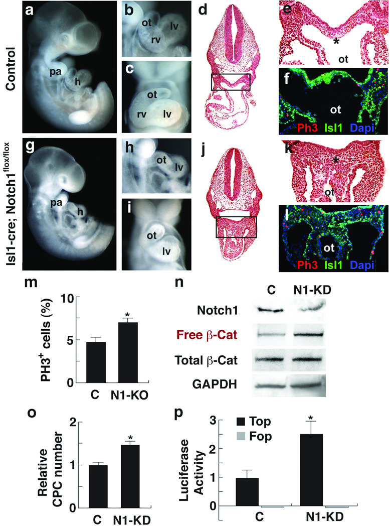
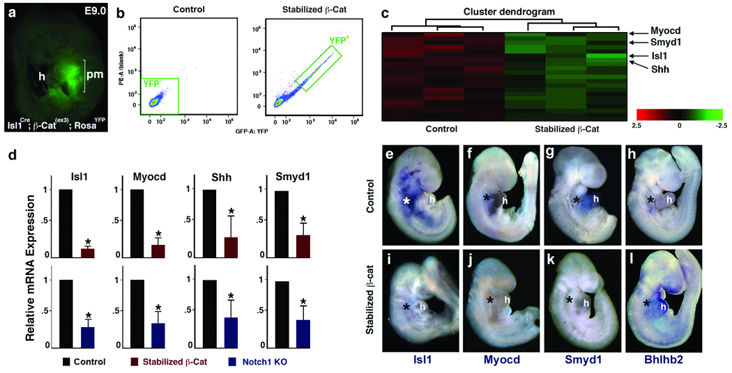
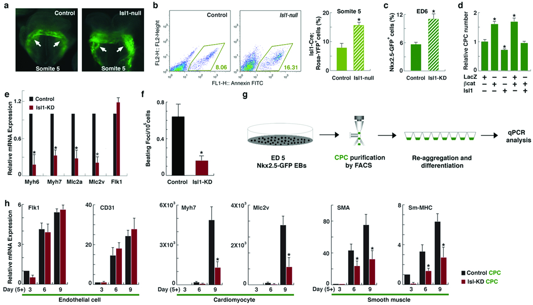
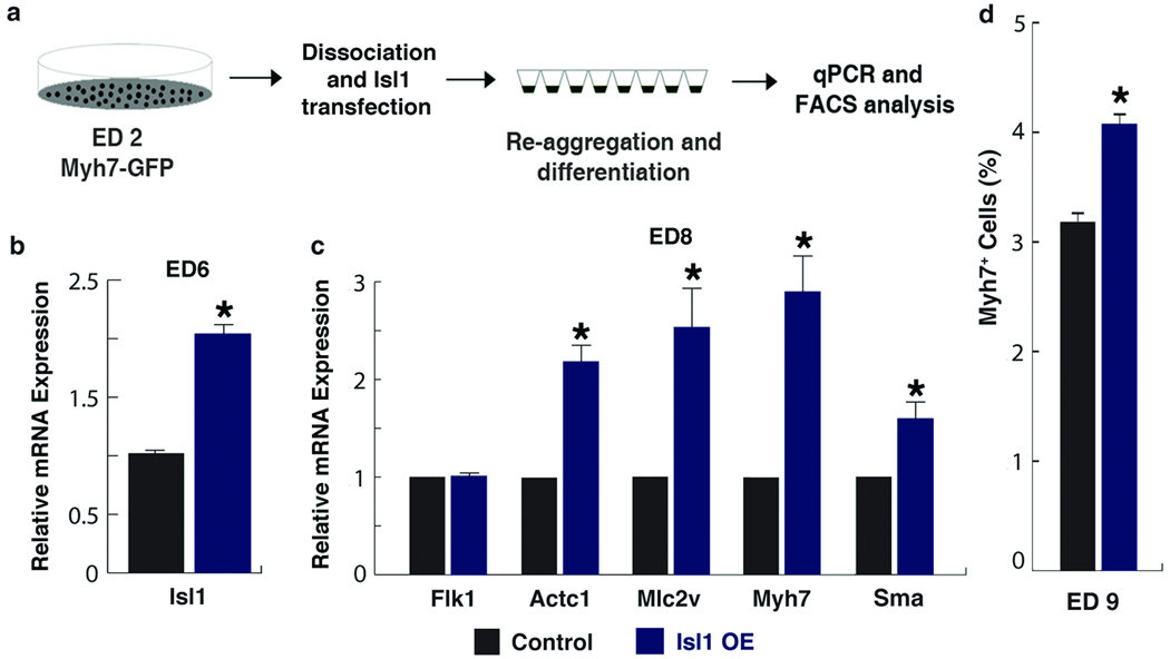
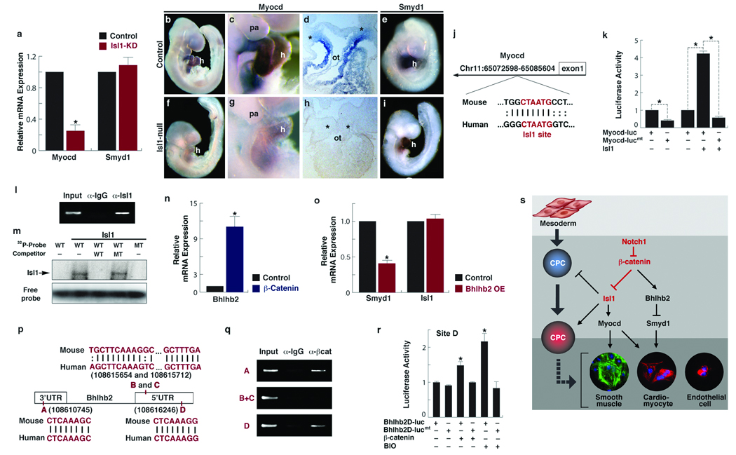
Regulation of multipotent cardiac progenitor cell (CPC) expansion and subsequent differentiation into cardiomyocytes, smooth muscle or endothelial cells is a fundamental aspect of basic cardiovascular biology and cardiac regenerative medicine. However, the mechanisms governing these decisions remain unclear. Here, we show that Wnt/beta-catenin signalling, which promotes expansion of CPCs, is negatively regulated by Notch1-mediated control of phosphorylated beta-catenin accumulation within CPCs, and that Notch1 activity in CPCs is required for their differentiation. Notch1 positively, and beta-catenin negatively, regulated expression of the cardiac transcription factors, Isl1, Myocd and Smyd1. Surprisingly, disruption of Isl1, normally expressed transiently in CPCs before their differentiation, resulted in expansion of CPCs in vivo and in an embryonic stem (ES) cell system. Furthermore, Isl1 was required for CPC differentiation into cardiomyocyte and smooth muscle cells, but not endothelial cells. These findings reveal a regulatory network controlling CPC expansion and cell fate that involves unanticipated functions of beta-catenin, Notch1 and Isl1 that may be leveraged for regenerative approaches involving CPCs.


Figures

Figure 1
Figure 1
Notch1 loss-of-function causes CPC expansion and increases free β-Catenin levels. a–f, Control embryos. g–l, Isl1Cre, Notch1flox/flox embryos (N1-KO). a,g, Lateral views of ED10.5 embryos. b, c, h, i, Lateral (b,h) or frontal (c,i) view of embryos focused on cardiac regions showing absence of right ventricle (rv) in mutants. d,e, j, k, Transverse sections (H&E) of embryos (d, j) with enlargement of boxed areas (e, k) showing hyperplasia of precardiac progenitors (asterisk). f, l, Phosphohistone3 (Ph3, red) and Isl1 (green) immunostaining of transverse sections through the precardiac region. To compensate for the severe downregulation of Isl1 in Notch1 mutant embryos, Isl1 signals were amplified with the TSA system. DAPI (blue) was used to counterstain the nuclei. m, Percentage of ph3-positive cells in precardiac mesoderm region shown in e and k (mean ± s. d.; n=4; *P < 0.01). n, Western analyses of FACS-purified CPCs transfected with control siRNA (C) or Notch1 siRNA (N1-KD) using Notch1, free or total β-Catenin antibodies. Free β-Catenin antibodies detect dephosphorylated β-Catenin, the effector molecule of the Wnt/β-Catenin signaling pathway. GAPDH antibody was used as a control. o, Relative number of cells on the 2nd day after transfecting CPCs with control or Notch1 siRNA (mean ± s. d.; n=6; *P < 0.01). p, Top/Fop flash activity in CPCs transfected with indicated siRNA. Top flash is a luciferase reporter with Tcf binding sites to read Wnt/β-Catenin signaling activity. Fop flash contains mutated Tcf binding sites. Luciferase values were normalized to Renilla activity (mean ± s. d.; n=3; *P < 0.01). h, heart; pa, pharyngeal arch; ot, outflow tract; lv, left ventricle. Scale bars, 250 µm (a, g) or 100 µm (b–e, h–k).
Figure 2
Figure 2
Identification of genes affected by stabilized β-Catenin in cardiac progenitors. a, Lateral view of RosaYFP; Isl1Cre; β-catenin(ex3)loxP embryo at E9.0 showing YFP+ cells in precardiac mesoderm (pm). b, Histograms of YFP+ cell populations from control (Isl1Cre, left) and stabilized β-cat (Isl1Cre; β-catenin(ex3)loxP, right) embryos. c, A heatmap of expression arrays showing significantly downregulated cardiac genes (green) in stabilized β-catenin pm cells. Color bar indicates fold change in log2 scale. d, qPCR data of downregulated genes in FACS-purified cardiac progenitors with stabilized β-Catenin (Top). These genes were similarly affected in pm of Notch1 loss-of-function embryos (Bottom). Data are mean ± s. d.; n=3; *P < 0.01. l, Whole-mount in situ hybridization of genes indicated from control (top) and stabilized β-Catenin (bottom) embryos at E 9.5. Asterisks indicate precardiac mesoderm. h, heart. Scale bars, 100 µm.
Figure 3
Figure 3
Isl1 loss-of-function results in expansion of CPCs and suppression of their myocardial and smooth muscle lineages.a, YFP expression in control (RosaYFP, Isl1Cre/+, left) and Isl1-null (RosaYFP, Isl1Cre/Cre, right) embryos at the 5-somite stage. Arrows indicate YFP+ CPCs. Scale bars, 50 µm. b, Quantification of YFP+ cells in indicated embryos at somite 5 (mean ± s. d.; n=3; *P < 0.01). c, Quantification of GFP+ cells in ED6 Nkx2.5-GFP EBs with or without Isl1 KD (mean ± s. d.; n=3; *P < 0.01). d, Relative number of cells on the 2nd day after transfecting EB-derived CPCs with lacZ, β-catenin, or Isl1 (mean ± s. d.; n=6; *P < 0.01). e, Relative mRNA expression of indicated genes in control or Isl1-KD EBs at ED 9, determined by qPCR (mean ± s. d.; n=4; *P < 0.01). f, Number of beating foci per 10 cells in control or Isl1-KD EBs at ED12. g, Schematic diagram of isolating CPCs from ES cells and their differentiation. h, Relative mRNA expression of endothelial (Flk1, CD31), cardiomyocyte (Myh7, Mlc2v) or smooth muscle (Sma, Sm-mhc) genes during CPC differentiation, determined by qPCR (mean ± s. e. m. ; n=4; *P < 0.05).
Figure 4
Figure 4
Increased levels of Isl1 promote myocardial differentiation. a, Schematic diagram of differentiation of Myh7-GFP ES cells with Isl1 overexpression. b, c, Relative expression levels of Isl1 on ED6 EBs (b), and endothelial (Flk1), cardiac sarcomeric (Actc1, Mlc2v, Myh7) and smooth muscle (Sma) genes on day 8 EBs (c), determined by qPCR. d, FACS analyses on ED 9 EBs to identify % of cells entering myocardial-lineage. Data are mean ± s. e. m.; n=3; *P < 0.005.
Figure 5
Figure 5
Isl1 targets Myocd and β-Catenin regulates Bhlhb2 to repress Smyd1. a, Relative expression levels of Myocd and Smyd1 in FACS-purified control and Isl1 knockdown (KD) CPCs, determined by qPCR (mean ± s. d.; n=4; *P < 0.005). b–i, Control (b–e) and Isl1-null (f–i) embryos at E 9.5 after in situ hybridization with Myocd (b–d, f–h), or Smyd1 (e, i) riboprobes. c, g, Lateral views focused on heart (h) and pharyngeal arch (pa) regions. d, h, Transverse section through the outfow tract. Asterisks indicate pre-cardiac mesoderm. Scale bars, 100 µm. j, Location of the conserved island containing Isl1 binding site (red) in the Myocd locus. k, Relative luciferase activity determined with luciferase reporters linked to the conserved island with the intact Isl1 site (Myocd-luc) or with a mutant Isl1 site (Myocd-lucmt) in the presence or absence of Isl1 (mean ± s. d.; n=3; *P < 0.005). l, Chromatin immunoprecipitation (ChIP) assay shows specific PCR amplification of the Isl1 consensus site shown in j, representing association with Isl1 protein. m, Electophoretic mobility shift assay with in-vitro synthesized Isl1 protein and radiolabeled probes (Probe) spanning the Isl1 site shown in j. Unlabeled probes were used as competitors. WT, wildtype; MT, mutant. n, Relative expression levels of Bhlhb2 in CPCs with stabilized β-Catenin, determined by qPCR (mean ±s. d.; n=3; *P < 0.005).. o, Relative expression levels of Smyd1 and Isl1 after transfecting FACS-purified CPCs with Bhlhb2 and differentiating them for 3 days (mean ± s. d.; n=3; *P < 0.005). p, The Bhlhb2 locus showing four conserved Lef/Tcf binding sites. q, ChIP assays performed with Lef/Tcf consensus sites shown in p. β-Catenin forms complexes with sites A and D as revealed by amplification of those sites. r, Relative luciferase activity determined with luciferase reporters containing the intact Lef/Tcf site D (Bhlhb2D-luc) or with a mutant Lef/Tcf site D (Bhlhb2D-lucmt) in the presence or absence of β-Catenin or BIO (2µM). Data are mean ± s. d.; n=3; *P < 0.005. s, A molecular cascade involving Notch1/ β-Catenin/ Isl1 during CPC fate determination. Notch1 functions to negatively regulate accumulation of free β-Catenin, which regulates Myocd and Smyd1 through Isl1 and Bhlhb2, respectively, to determine CPC fates. Relationships indicated may be direct or indirect.

References

    1. Cohen ED, et al. Wnt/beta-catenin signaling promotes expansion of Isl-1-positive cardiac progenitor cells through regulation of FGF signaling. J Clin Invest. 2007;117:1794–1804. - PMC - PubMed
    1. Kwon C, et al. Canonical Wnt signaling is a positive regulator of mammalian cardiac progenitors. Proc Natl Acad Sci U S A. 2007;104:10894–10899. - PMC - PubMed
    1. Qyang Y, et al. The renewal and differentiation of Isl1+ cardiovascular progenitors are controlled by a Wnt/beta-catenin pathway. Cell Stem Cell. 2007;1:165–179. - PubMed
    1. Cai CL, et al. Isl1 identifies a cardiac progenitor population that proliferates prior to differentiation and contributes a majority of cells to the heart. Dev Cell. 2003;5:877–889. - PMC - PubMed
    1. Yang L, et al. Human cardiovascular progenitor cells develop from a KDR+ embryonic-stem-cell-derived population. Nature. 2008;453:524–528. - PubMed

Publication types

MeSH terms

Associated data