. 2009 Dec;41(12):1350-3.
doi: 10.1038/ng.471. Epub 2009 Nov 1.

Differential methylation of tissue- and cancer-specific CpG island shores distinguishes human induced pluripotent stem cells, embryonic stem cells and fibroblasts

Affiliations

Differential methylation of tissue- and cancer-specific CpG island shores distinguishes human induced pluripotent stem cells, embryonic stem cells and fibroblasts

Akiko Doi et al. Nat Genet. 2009 Dec.

Abstract

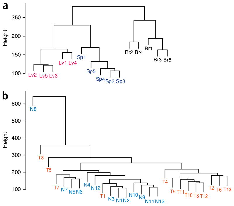
Induced pluripotent stem (iPS) cells are derived by epigenetic reprogramming, but their DNA methylation patterns have not yet been analyzed on a genome-wide scale. Here, we find substantial hypermethylation and hypomethylation of cytosine-phosphate-guanine (CpG) island shores in nine human iPS cell lines as compared to their parental fibroblasts. The differentially methylated regions (DMRs) in the reprogrammed cells (denoted R-DMRs) were significantly enriched in tissue-specific (T-DMRs; 2.6-fold, P < 10(-4)) and cancer-specific DMRs (C-DMRs; 3.6-fold, P < 10(-4)). Notably, even though the iPS cells are derived from fibroblasts, their R-DMRs can distinguish between normal brain, liver and spleen cells and between colon cancer and normal colon cells. Thus, many DMRs are broadly involved in tissue differentiation, epigenetic reprogramming and cancer. We observed colocalization of hypomethylated R-DMRs with hypermethylated C-DMRs and bivalent chromatin marks, and colocalization of hypermethylated R-DMRs with hypomethylated C-DMRs and the absence of bivalent marks, suggesting two mechanisms for epigenetic reprogramming in iPS cells and cancer.


Figures

Figure 1
Figure 1
Reprogramming differentially methylated regions (R-DMRs). (a) Enrichment of R-DMRs at CpG island shores. The CHARM array (left, labeled CpG regions) is enriched in CpG islands, and the R-DMRs (right, labeled R-DMR) show marked enrichment at CpG island shores. Islands are denoted as regions that include >50% of a CpG island or are wholly contained in an island, and overlap regions are denoted as regions that include 0.1–50% of a CpG island. Specific base intervals of regions not overlapping islands are indicated; (0–500) means from 1 to 500 bases. Percentage of the distribution (y axis) is given for the CpG regions (CHARM array, null hypothesis) and reprogramming differentially methylated regions (R-DMRs). (b,c) Examples of DMRs. The gene encoding bone morphogenetic protein 7 (BMP7) is indicated in b, and the gene encoding goosecoid (GSC) is indicated in c. In each case, the upper panels show a plot of methylation (M value; see Online Methods) versus genomic location, where the curve represents averaged smoothed M values; the location of CpG dinucleotides (black tick marks), CpG density, location of CpG islands (orange line), as well as the gene annotation are shown. The bottom panels show validation by bisulfite pyrosequencing (mapping to red box in upper panel). Bars represent the mean methylation (triplicate measurement) ± s.d. of iPS cells (pink), fibroblasts (gray) and ES cells (blue) as well as the generally highly methylated HCT116 colon cancer cell line and a generally hypomethylated double DNA methyltransferase 1/3B double knockout line (DKO) derived from it. In each case, five separate CpG sites were assayed quantitatively, shown as differing shades.
Figure 2
Figure 2
DNA methylation at R-DMRs distinguishes normal tissues from each other and colon cancer from normal colon. (a,b) The M values of all tissues from the 4,401 regions (FDR < 0.05) corresponding to R-DMRs (iPS cells compared to parental fibroblasts) were used for unsupervised hierarchical clustering comparing (a) normal brain, spleen and liver (denoted as Br, Sp and Lv, respectively) and (b) colorectal cancer and matched normal colonic mucosa (denoted as T and N, respectively). Notably, all of the normal brain, spleen and liver tissues are completely discriminated by the regions that differ between iPS cells and fibroblasts (R-DMRs). The major branches in the dendrograms correspond perfectly to tissue type. Furthermore, most of the colorectal cancer samples are discriminated from matched normal colonic mucosa by R-DMRs.

References

    1. Takahashi K, Yamanaka S. Induction of pluripotent stem cells from mouse embryonic and adult fibroblast cultures by defined factors. Cell. 2006;126:663–676. - PubMed
    1. Takahashi K, et al. Induction of pluripotent stem cells from adult human fibroblasts by defined factors. Cell. 2007;131:861–872. - PubMed
    1. Park IH, et al. Reprogramming of human somatic cells to pluripotency with defined factors. Nature. 2008;451:141–146. - PubMed
    1. Yu J, et al. Induced pluripotent stem cell lines derived from human somatic cells. Science. 2007;318:1917–1920. - PubMed
    1. Park IH, et al. Disease-specific induced pluripotent stem cells. Cell. 2008;134:877–886. - PMC - PubMed

Publication types

Associated data