. 2011 Feb 22;108(8):3336-41.
doi: 10.1073/pnas.1012351108. Epub 2011 Feb 2.

RNAi screen of the protein kinome identifies checkpoint kinase 1 (CHK1) as a therapeutic target in neuroblastoma

Affiliations

RNAi screen of the protein kinome identifies checkpoint kinase 1 (CHK1) as a therapeutic target in neuroblastoma

Kristina A Cole et al. Proc Natl Acad Sci U S A. .

Abstract

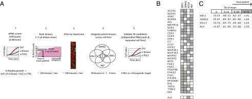
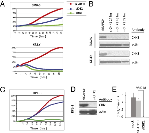
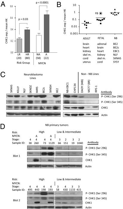
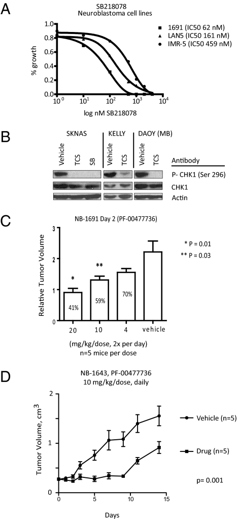
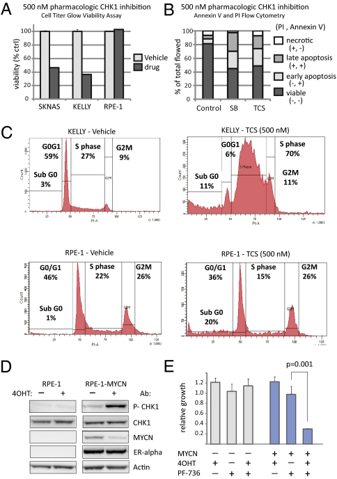
Neuroblastoma is a childhood cancer that is often fatal despite intense multimodality therapy. In an effort to identify therapeutic targets for this disease, we performed a comprehensive loss-of-function screen of the protein kinome. Thirty kinases showed significant cellular cytotoxicity when depleted, with loss of the cell cycle checkpoint kinase 1 (CHK1/CHEK1) being the most potent. CHK1 mRNA expression was higher in MYC-Neuroblastoma-related (MYCN)-amplified (P < 0.0001) and high-risk (P = 0.03) tumors. Western blotting revealed that CHK1 was constitutively phosphorylated at the ataxia telangiectasia response kinase target site Ser345 and the autophosphorylation site Ser296 in neuroblastoma cell lines. This pattern was also seen in six of eight high-risk primary tumors but not in control nonneuroblastoma cell lines or in seven of eight low-risk primary tumors. Neuroblastoma cells were sensitive to the two CHK1 inhibitors SB21807 and TCS2312, with median IC(50) values of 564 nM and 548 nM, respectively. In contrast, the control lines had high micromolar IC(50) values, indicating a strong correlation between CHK1 phosphorylation and CHK1 inhibitor sensitivity (P = 0.0004). Furthermore, cell cycle analysis revealed that CHK1 inhibition in neuroblastoma cells caused apoptosis during S-phase, consistent with its role in replication fork progression. CHK1 inhibitor sensitivity correlated with total MYC(N) protein levels, and inducing MYCN in retinal pigmented epithelial cells resulted in CHK1 phosphorylation, which caused growth inhibition when inhibited. These data show the power of a functional RNAi screen to identify tractable therapeutical targets in neuroblastoma and support CHK1 inhibition strategies in this disease.


Conflict of interest statement

The authors declare no conflict of interest.

Figures

Fig. 1.
Fig. 1.
RNAi screen in neuroblastoma reveals a list of candidate kinase targets. (A) Schematic of the RNAi kinase screen in neuroblastoma. (1) Four neuroblastoma cell lines were treated with 529 kinase siRNAs and analyzed for substrate adherent growth. (2) Most active kinases of each cell line were ranked if the percentage of relative growth (AUC kinase/AUCctrl) was less than 0.5 SD of the mean percentage of relative growth for that line. (3) Kinases were further filtered for expression within their line. (4) Integration of the most potent kinases. (B) Thirty candidate kinases emerged as targets in three or more cell lines in the screen. ALK emerged as a candidate kinase in KELLY, the only ALK mutated line in the screen. (C) The percentage of relative growth of each cell line was divided by quartile within a cell line and represented as progressively darker boxes by increasing potency.
Fig. 2.
Fig. 2.
CHK1 depletion was the most potent inhibitor of neuroblastoma growth. (A) Growth curves of neuroblastoma cell lines SKNAS and KELLY treated with siCHK1 and siGAPDH and the positive control siPLK1. (B) Western blot of siCHK protein depletion in KELLY and SKNAS. CHK1 depletion does not significantly inhibit the growth of control RPE1 cells (C) despite adequate mRNA (D) and protein depletion (E).
Fig. 3.
Fig. 3.
CHK1 mRNA and protein are highly expressed in neuroblastoma. (A) mRNA expression profiling of 100 neuroblastoma diagnostic tumors showed that CHK1 is overexpressed in HR tumors compared with LR tumors (Left) and in MYCN-amplified tumors compared with MYCN NA tumors (Right) (mean ± SEM). HR, high-risk; LR, low-risk; NA, single-copy. (B) CHK1 mRNA expression by real-time PCR demonstrates that CHK1 is overexpressed in neuroblastoma cell lines and embryonal tissues compared with adult tissues. (C) Western blot with constitutive phosphorylation of CHK1 Ser296 and Ser345 in neuroblastoma cell lines compared with control cell lines. (D) Western blot of phosphorylation of CHK1 Ser296 and Ser345 and CHK1 in neuroblastoma primary tumors. A, genomic amplification of MYCN (samples 260 and 1,129). Each of the two blots (blot 1 and blot 2) had four low/intermediate-risk and four high-risk tumors run in parallel. Fig. S6 shows the original order of blot 1, and blot 2 is ordered as in the figure.
Fig. 4.
Fig. 4.
Pharmacologic inhibition of CHK1 is potent in neuroblastoma cells with activated CHK1. (A) Three dose–response curves of SB in neuroblastoma cell lines. (B) Western blots of CHK1 Ser296 from KELLY, SKNAS, and DAOY cells treated with 500 nM TCS or SB for 48 h. (C) Five mice per cohort bearing NB1691 xenografts are treated with vehicle or with the CHK1 inhibitor PF-00477736 at a dose of 4, 10, or 20 mg/kg twice daily, and relative tumor volumes are determined on day 2 (mean ± SEM). (D) Mice bearing NB1643 xenografts are treated daily with 10 mg/kg of the CHK1 inhibitor PF-00477736 for 14 d, and tumor volumes are shown (mean ± SEM).
Fig. 5.
Fig. 5.
CHK1 is phosphorylated in response to MYCN activation, and CHK1 inhibition causes apoptosis and replication arrest. (A) Treatment of the neuroblastoma cell lines SKNAS and KELLY with the CHK1 inhibitor SB (500 nM) decreased cellular viability by >50% (average ± SD; P < 0.0001), whereas the control RPE-1 cells are viable. (B) CHK1 inhibition by TCS and SB caused significant apoptosis in KELLY cells by 48 h as determined by annexin/PI staining. (C) Representative cell cycle histogram by PI staining demonstrated that CHK1 inhibited by TCS in neuroblastoma KELLY cells harvested at 48 h after treatment are arrested during replication compared with control (P < 0.0001), whereas there is no S-phase arrest in RPE1 cells. (D) Western blot of CHK1 Ser296, total CHK1, MYCN-ER (as analyzed by an anti-MYCN and an anti-ER antibody, 90 kDa), and actin in RPE1–MYCN-ER or parental RPE1 cells with and without MYCN-ER activation by 4-OHT. (E) There is a significant decrease (P = 0.001) in cellular growth after CHK1 inhibition by PF-00477736 (PF-736) only in RPE1 cells with activated MYCN (RPE1–MYCN-ER treated with 4-OHT).

References

    1. Maris JM. Recent advances in neuroblastoma. N Engl J Med. 2010;362:2202–2211. - PMC - PubMed
    1. Matthay KK, et al. Role of myeloablative therapy in improved outcome for high risk neuroblastoma: Review of recent Children's Cancer Group results. Eur J Cancer. 1995;31A:572–575. - PubMed
    1. Mossé YP, et al. Identification of ALK as a major familial neuroblastoma predisposition gene. Nature. 2008;455:930–935. - PMC - PubMed
    1. George RE, et al. Activating mutations in ALK provide a therapeutic target in neuroblastoma. Nature. 2008;455:975–978. - PMC - PubMed
    1. Molenaar JJ, et al. Inactivation of CDK2 is synthetically lethal to MYCN over-expressing cancer cells. Proc Natl Acad Sci USA. 2009;106:12968–12973. - PMC - PubMed

Publication types

Associated data