. 2010 Aug 3;107(31):13794-9.
doi: 10.1073/pnas.1008684107. Epub 2010 Jul 16.

NLR family member NLRC5 is a transcriptional regulator of MHC class I genes

Affiliations

NLR family member NLRC5 is a transcriptional regulator of MHC class I genes

Torsten B Meissner et al. Proc Natl Acad Sci U S A. .

Abstract

MHC class I plays a critical role in the immune defense against viruses and tumors by presenting antigens to CD8 T cells. An NLR protein, class II transactivator (CIITA), is a key regulator of MHC class II gene expression that associates and cooperates with transcription factors in the MHC class II promoter. Although CIITA also transactivates MHC class I gene promoters, loss of CIITA in humans and mice results in the severe reduction of only MHC class II expression, suggesting that additional mechanisms regulate the expression of MHC class I. Here, we identify another member of the NLR protein family, NLRC5, as a transcriptional regulator of MHC class I genes. Similar to CIITA, NLRC5 is an IFN-gamma-inducible nuclear protein, and the expression of NLRC5 resulted in enhanced MHC class I expression in lymphoid as well as epithelial cell lines. Using chromatin immunoprecipitation and reporter gene assays, we show that NLRC5 associates with and activates the promoters of MHC class I genes. Furthermore, we show that the IFN-gamma-induced up-regulation of MHC class I requires NLRC5, because knockdown of NLRC5 specifically impaired the expression of MHC class I. In addition to MHC class I genes, NLRC5 also induced the expression of beta2-microglobulin, transporter associated with antigen processing, and large multifunctional protease, which are essential for MHC class I antigen presentation. Our results suggest that NLRC5 is a transcriptional regulator, orchestrating the concerted expression of critical components in the MHC class I pathway.


Conflict of interest statement

The authors declare no conflict of interest.

Figures

Fig. 1.
Fig. 1.
NLRC5 contains an N-terminal bipartite NLS and can translocate into the nucleus. (A, D, and F) HEK293T cells were transfected with expression plasmids coding for GFP or the indicated GFP fusion proteins; 48 h posttransfection, cells were treated with 10 nM leptomycin B (LMB) for 90 min or left untreated. Fixed cells were stained with Hoechst 33342 to indicate the nuclei. (Scale bar: 10 μm.) (A) Cellular localization of NLRC5 and CIITA on LMB treatment. (B) Phylogenetic tree of CARD-containing NLRs. (C) Schematic representation of the NLRC5 deletion mutant constructs used to map the nuclear localization signal. The position of the NLS is indicated by an asterisk. (D) Cellular localization of NLRC5 deletion mutants on LMB treatment. (E) Sequence of the bipartite NLS found in the N terminus of NLRC5. Alanine substitution of the right or left arm of the NLS was used to construct the NLSI and NLSII import mutant-expression plasmids. (F) Cellular localization of the NLSI and NLSII mutant forms of NLRC5 on LMB treatment.
Fig. 2.
Fig. 2.
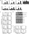
Induction of MHC class I and functionally related genes by NLRC5. (A) RNA isolated from Jurkat T cells stably expressing the indicated GFP fusion proteins was analyzed by quantitative real-time PCR for the expression of the indicated genes. GFP, empty vector; WT, wild-type NLRC5; A, Walker A mutant; B, Walker B mutant; AB, Walker AB mutant. (B) The same Jurkat T cell lines were examined for the expression of MHC class I heavy chain (HC), β2M, TAP1, and LMP2 by Western blot analysis. Actin levels are shown as a loading control. (C) Surface expression of MHC class I in Jurkat T cell lines expressing GFP (gray line) or the indicated GFP-NLRC5 fusion proteins (black line) was examined by flow cytometry using anti–pan-MHC class I (HLA-A, -B, and -C) and HLA-E antibodies. IFN-γ (100 U/mL) treatment was used as a positive control. Data obtained with an isotype control antibody are indicated by the shaded area. (D) HEK293T cells were transiently transfected with the expression plasmids for GFP fused to NLRC5, CIITA (black line), or GFP only (gray line). The expression of MHC class I (HLA-A, -B, and -C) or class II (HLA-DR) was analyzed by flow cytometry 48 h posttransfection. Data obtained with an isotype control antibody are indicated by the shaded area.
Fig. 3.
Fig. 3.
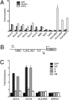
NLRC5 binds and transactivates MHC class I gene promoters. (A) NLRC5-mediated transactivation of MHC class I and functionally related genes. HEK293T cells were transiently transfected with either expression vectors for GFP, GFP-NLRC5, or GFP-CIITA, along with luciferase reporter constructs of the indicated gene promoters. Cell lysates were analyzed 48 h posttransfection by dual-luciferase assay. Data are a representative of three independent experiments performed in duplicates, and error bars represent ± SD. (B) Schematic representation of the W/SXY module found in the promoters of MHC class I and class II genes. The position of the primers used in the ChIP assay is indicated with arrows (P1 and P2). (C) NLRC5 occupancy, in terms of fold enrichment, at the HLA-A, -B, or -DRA promoters as determined by ChIP. Jurkat T cells stably expressing the indicated GFP fusion proteins were analyzed by ChIP assay using an anti-GFP antibody for immunoprecipitation and the indicated qPCR primers (B). GFP, empty vector; WT, wild-type NLRC5; A, Walker A mutant; B, Walker B mutant; AB, Walker AB mutant. Error bars indicate SEM (± SEM) from three independent experiments.
Fig. 4.
Fig. 4.
Knockdown of NLRC5 results in decreased up-regulation of MHC class I on IFN-γ treatment. (A) HeLa cells were stimulated with IFN-γ (100 U/mL) for the indicated time points, and the kinetics of NLRC5, HLA-A, and STAT1 expression were analyzed by quantitative real-time PCR. (B and C) HeLa cells were transfected with NLRC5-specific or control siRNAs; 16 h posttransfection, cells were stimulated with IFN-γ for 24 h. (B) Knockdown efficiency of NLRC5 was determined by quantitative real-time PCR using gene-specific primers, and data were normalized to the expression of the GAPDH gene. Scr, control scrambled siRNA. Error bars represent the ± SD from one representative of three independent experiments performed in duplicates. *P < 0.05. (C) Surface expression of MHC class I and β1-integrin was analyzed by flow cytometry. (D) Model depicting the role of NLRC5 in the IFN-γ–induced up-regulation of MHC class I genes. Discussion has further details.

References

    1. Pamer E, Cresswell P. Mechanisms of MHC class I—restricted antigen processing. Annu Rev Immunol. 1998;16:323–358. - PubMed
    1. Braud VM, Allan DS, McMichael AJ. Functions of nonclassical MHC and non-MHC-encoded class I molecules. Curr Opin Immunol. 1999;11:100–108. - PubMed
    1. Le Bouteiller P, Solier C. Is antigen presentation the primary function of HLA-G? Microbes Infect. 2001;3:323–332. - PubMed
    1. Shastri N, Cardinaud S, Schwab SR, Serwold T, Kunisawa J. All the peptides that fit: The beginning, the middle, and the end of the MHC class I antigen-processing pathway. Immunol Rev. 2005;207:31–41. - PubMed
    1. Peaper DR, Cresswell P. Regulation of MHC class I assembly and peptide binding. Annu Rev Cell Dev Biol. 2008;24:343–368. - PubMed

Publication types

MeSH terms

Substances

Associated data