. 2023 Nov 20;14(1):7555.
doi: 10.1038/s41467-023-43369-x.

Protective effects of macrophage-specific integrin α5 in myocardial infarction are associated with accentuated angiogenesis

Affiliations

Protective effects of macrophage-specific integrin α5 in myocardial infarction are associated with accentuated angiogenesis

Ruoshui Li et al. Nat Commun. .

Abstract

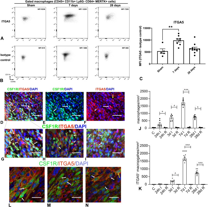
Macrophages sense changes in the extracellular matrix environment through the integrins and play a central role in regulation of the reparative response after myocardial infarction. Here we show that macrophage integrin α5 protects the infarcted heart from adverse remodeling and that the protective actions are associated with acquisition of an angiogenic macrophage phenotype. We demonstrate that myeloid cell- and macrophage-specific integrin α5 knockout mice have accentuated adverse post-infarction remodeling, accompanied by reduced angiogenesis in the infarct and border zone. Single cell RNA-sequencing identifies an angiogenic infarct macrophage population with high Itga5 expression. The angiogenic effects of integrin α5 in macrophages involve upregulation of Vascular Endothelial Growth Factor A. RNA-sequencing of the macrophage transcriptome in vivo and in vitro followed by bioinformatic analysis identifies several intracellular kinases as potential downstream targets of integrin α5. Neutralization assays demonstrate that the angiogenic actions of integrin α5-stimulated macrophages involve activation of Focal Adhesion Kinase and Phosphoinositide 3 Kinase cascades.


Conflict of interest statement

The authors declare no competing interests.

Figures

Fig. 1
Fig. 1. Time course of ITGA5 expression in infarct macrophages.
AC Flow cytometry was used to assess ITGA5 expression in infarct macrophages. The gating strategy is shown in Supplementary Fig. 2. Representative images show the mean fluorescent intensity (MFI) for ITGA5 antibody (A) and isotype control (B) in CD45 + /CD11b + /Ly6G-/CD64 + /MerTK+ macrophages in sham animals and in infarcts. Quantitative analysis showed a 2-fold increase in ITGA5 mean fluorescent intensity (MFI) in macrophages 7 days after infarction (**p < 0.01; Sham: n = 5 biologically independent experiments, 7-day: n = 8 biologically independent experiments, 28-day: n = 11 biologically independent experiments). Statistical analysis was performed using one-way ANOVA followed by Bonferroni post-hoc test. To examine the time course of ITGA5 expression in infarct macrophages, we have also performed dual immunofluorescence in myocardial sections from control and infarcted CSF1REGFP macrophage reporter mice, combining ITGA5 staining (red) and CSF1R (GFP) labeling. Abundant ITGA5+ macrophages were noted in 7-day infarcts (DI, arrows). CSF1R-negative cells with vascular, or fibroblast morphology (DF, yellow arrow) also expressed ITGA5. J Quantitative analysis showed that the density of CSF1R+ macrophages was significantly higher in infarcted segments, when compared with remote non-infarcted myocardium from the same timepoint (^p < 0.05, ^^^p < 0.001, n = 6 biologically independent experiments in control (C), 24-hour, 7-day and 28-day groups, n = 5 biologically independent experiments in the 3-day group). Statistical analysis was performed using non-parametric ANOVA (Kruskal-Wallis) followed by Dunn’s post-hoc test. No significant increase in the density of myeloid cells was noted in non-infarcted remodeling segments. K An increase in the density of ITGA5+ macrophages was first noted 3 days after coronary occlusion, and peaked after 7 days of coronary occlusion, but was significantly reduced at the 28-day timepoint (*p < 0.05, **p < 0.01 ****p < 0.0001 vs. C ^p < 0.05, ^^^p < 0.001, vs. corresponding remote non-infarcted myocardium, n = 6 biologically independent experiments in control (C), 24-hour, 7-day and 28-day groups, n = 5 biologically independent experiments in 3-day group). Statistical analysis was performed using non-parametric ANOVA (Kruskal–Wallis) followed by Dunn’s post-hoc test. LN Confocal microscopy showed that ITGA5 was localized not only on the macrophage surface (white arrows), but also in the cytoplasm (yellow arrow), likely reflecting de novo synthesis of ITGA5, followed by subsequent shuttling to the cell membrane. Representative images were selected from 50 different scanned fields from 6 biologically independent experiments. Scalebar = 50 μm (for panels DF), =30 μm (panels GI), = 10 μm (panels LN). All data are shown as mean values +/- SEM. Source data are provided as a Source Data file.
Fig. 2
Fig. 2. Myeloid cell-specific ITGA5 loss accentuates dilative post-infarction remodeling.
Comparison of the survival curves between ITGA5 fl/fl and Myα5KO (myeloid cell–specific ITGA5 knockout) mice after 7-28 days of permanent coronary occlusion (AC). AC, no significant differences in mortality were noted between ITGA5 fl/fl and Myα5KO mice (ITGA5 fl/fl: n = 35, Myα5KO: n = 33) in male and female groups. Please note that male mice (B) have higher mortality than females (A) after myocardial infarction in both genotypes. Statistical analysis was performed using the log-rank test. DF Echocardiographic analysis showed that Myα5KO mice had accentuated dilative remodeling evidenced by increased left ventricular end-diastolic volume (LVEDV; D) and left ventricular end-systolic volume (LVESV; E) after 28 days. F: Although Myα5KO mice had a trend towards worse systolic dysfunction at the 28-day timepoint, the effects of myeloid cell-specific ITGA5 loss on left ventricular ejection fraction (LVEF; F) did not reach statistical significance (**p < 0.01, ITGA5 fl/fl pre: n = 26, Myα5KO pre: n = 24, ITGA5 fl/fl 7d: n = 12, Myα5KO 7d: n = 11, ITGA5 fl/fl 28d: n = 13, Myα5KO 28d: n = 13 biologically independent experiments). Statistical analysis was performed using one-way ANOVA, followed by the Sidak post-hoc test. Data are shown as mean values +/- SEM. Source data are provided as a Source Data file.
Fig. 3
Fig. 3. Myeloid cell-specific ITGA5 loss perturbs scar remodeling after myocardial infarction.
A In order to assess scar size after 7-28 days of coronary occlusion, systematic quantification of morphometric parameters was performed by sectioning and staining the entire heart from base to apex. B The area of infarcted myocardium was comparable between groups at the 7-day timepoint; however, Myα5KO mice exhibited significantly larger scars 28 days after infarction (*p < 0.05, ITGA5 fl/fl 7d: n = 14, Myα5KO 7d: n = 12, ITGA5 fl/fl 28d: n = 13, Myα5KO 28d: n = 13 biologically independent experiments). C, D Myα5KO mice had trends towards lower infarct wall thickness and larger infarct volume at the 28-day timepoint that did not reach statistical significance. Statistical analysis was performed using one-way ANOVA, followed by the Sidak post-hoc test. Data are shown as mean values +/- SEM. Source data are provided as a Source Data file. Scale bar = 2 mm.
Fig. 4
Fig. 4. Myeloid cell-specific ITGA5 loss does not affect infiltration of the infarcted heart with myeloid cells, macrophages, neutrophils, and T lymphocytes.
Flow cytometry was used to compare the percentage and absolute number of cells in the infarct, 7 days after myocardial infarction. The gating strategy is shown in Supplementary Fig. 7. DAPI was used to label live cells (DAPI-cells). Myeloid cells were identified as DAPI-/CD45 + /CD11b+ cells (A), macrophages were labeled as DAPI-/CD45 + /CD11b + /Ly6G-/CD64 + /MerTK+ cells (B), whereas T cells were identified as DAPI-/CD45 + /CD11b-/CD3e+ cells (C). Quantitative analysis showed no significant effects of myeloid cell-specific ITGA5 loss on myeloid cell (D, E), macrophage (F, G), neutrophil (H, I), and T cell (J, K) numbers in the infarct (p = NS, n = 5 biologically independent experiments/group). Panels D, F, H, and J show the percentage of each population in relation to all live cells (DAPI-cells), whereas panels E, G, I, and L show the absolute number of cells per mg of myocardial tissue. Data are shown as mean values +/- SEM, Statistical analysis was performed using unpaired two-tailed Student’s t test. Source data are provided as a Source Data file. NS no significance.
Fig. 5
Fig. 5. Myeloid cell-specific ITGA5 loss reduces microvascular density and perturbs formation of coated mature vessels in the healing infarct.
A CD31 immunohistochemistry was used to label endothelial cells. Representative images show CD31 staining of the infarcted area, border zone and remote remodeling myocardium from ITGA5 fl/fl and Myα5KO mice after 7 and 28 days of coronary occlusion. The arrow identifies a typical CD31+ microvessel. Quantitative analysis showed that Myα5KO mice have significantly reduced microvascular density in the infarct zone at the 7-day timepoint (B), and in the border zone (C) after 28 days of coronary occlusion. Microvascular density in the remote remodeling myocardium was comparable between Myα5KO mice and corresponding ITGA5 fl/fl controls (D) (**p < 0.01, ***p < 0.001, ITGA5 fl/fl 7d: n = 14, Myα5KO 7d: n = 12, ITGA5 fl/fl 28d: n = 13, Myα5KO 28d: n = 13 biologically independent experiments). Scalebar = 100um. E In healing infarcts, microvessels undergo maturation acquiring a coat comprised of α-SMA-expressing mural cells (arrowheads). Representative images show α-SMA immunofluorescence staining of infarcted and remote areas from ITGA5 fl/fl and Myα5KO mice after 7 and 28 days of coronary occlusion. The arrow identifies a typical α-SMA+ vessel. Myα5KO mice had significantly reduced density of mature α-SMA+ microvessels in the infarct zone at the 28-day timepoint (F). The density of α-SMA+ vessels in the remote remodeling myocardium was comparable between Myα5KO and ITGA5 fl/fl mice (G). (****p < 0.0001, ITGA5 fl/fl 7d: n = 14, Myα5KO 7d: n = 12, ITGA5 fl/fl 28d: n = 13, Myα5KO 28d: n = 13 biologically independent experiments). Data are shown as mean values +/- SEM. Statistical analysis was performed using one-way ANOVA, followed by Sidak post-hoc test. Source data are provided as a Source Data file. Scalebar = 100um. I infarct area, R remote area.
Fig. 6
Fig. 6. Conditional ITGA5 deletion in CX3CR1+ macrophages accentuates dilative post-infarction remodeling, perturbing infarct angiogenesis and vascular maturation.
Echocardiographic assessment showed that inducible macrophage–specific ITGA5 knockout mice (iMaα5KO) had accentuated dilative remodeling after myocardial infarction, evidenced by increased left ventricular end-diastolic volume (LVEDV; A) and left ventricular end-systolic volume (LVESV; B) after 28 days of coronary occlusion. C The effects of macrophage-specific ITGA5 loss on left ventricular ejection fraction (LVEF; C) did not reach statistical significance (***p < 0.001, ITGA5 fl/fl pre: n = 14, iMaα5KO pre: n = 14, ITGA5 fl/fl 7d: n = 5, iMaα5KO 7d: n = 4, ITGA5 fl/fl 28d: n = 9, iMaα5KO 28d: n = 9 biologically independent experiments). D Representative images show CD31 immunohistochemical staining of infarcted area, border zone, and remote remodeling myocardium in ITGA5 fl/fl and iMaα5KO mice after 7 and 28 days of coronary occlusion. The arrow identifies a typical microvessel. iMaα5KO mice had significantly reduced microvascular density in the infarct zone (E) and in the border zone (F) after 28 days of coronary occlusion. Microvascular density in the remote remodeling myocardium was comparable between iMaα5KO mice and corresponding ITGA5 fl/fl controls (G). H In healing infarcts, microvessels undergo maturation acquiring a coat comprised of α-SMA-expressing mural cells (arrowheads). Representative images of α-SMA immunofluorescence staining from infarcted area and remote area of ITGA5 fl/fl and iMaα5KO mice after 7 and 28 days of coronary occlusions are shown. The arrow identifies a typical α-SMA+ microvessel. iMaα5KO mice had significantly reduced density of mature α-SMA+ microvessels in the infarct zone at the 28-day timepoint (I). The density of α-SMA+ vessels in the remote remodeling myocardium was comparable between groups (J) (****p < 0.0001, *p < 0.05, ITGA5 fl/fl 7d: n = 5, iMaα5KO 7d: n = 5, ITGA5 fl/fl 28d: n = 9, iMaα5KO 28d: n = 9 biologically independent experiments). Statistical analysis was performed using one-way ANOVA followed by post-hoc Sidak tests. Data are shown as mean values +/- SEM. Source data are provided as a Source Data file. Scale bar = 100um. I infarct area, R remote area.
Fig. 7
Fig. 7. Clusters of CSF1R+ cells in control and infarcted hearts.
ScRNA-seq was used to characterize the transcriptional profile of CSF1R+ cells harvested from control and infarcted hearts. 12 distinct clusters were identified (UMAP, A). B The heatmap illustrates relative expression of marker genes for various clusters. A cluster of reparative macrophages (RMp) was abundant in infarcted hearts after 7 days of coronary occlusion. Several additional macrophage clusters were identified (see text). In addition, cells with monocyte (Mo), granulocyte (Gr), and dendritic cell profiles (Dc) were also noted. A small cluster exhibited transcriptional characteristics of fibroblasts (Fib). C Relative expression of growth factors, inflammatory cytokines, matricellular genes, and proliferation-associated genes by various clusters. RMp and angiogenic macrophages (Amp) exhibited high levels of growth factor expression (blue arrows). In contrast, inflammatory macrophages (IMp) expressed high levels of inflammatory cytokines (red arrow). A cluster with high expression of proliferation-associated genes (such as Ccnb1, Ccne1, and Plk1) was also noted and may represent proliferative macrophages (PMp, green arrow). D Expression of angiogenic growth factors across macrophage clusters. Amp cells (which had high levels of Itga5) exhibited high expression of angiogenic genes (blue arrow). PMp proliferative macrophages, CMp resident cardiac macrophages, Mp macrophages. Source data are provided as a Source Data file. Raw scRNA-seq data were deposited in the NCBI’s Gene Expression Omnibus under accession number GSE227251.
Fig. 8
Fig. 8. Cluster-specific patterns of integrin expression in infarct macrophages.
A The heatmap shows the patterns of integrin gene expression across various clusters of CSF1R+ cells. B Although Itga5 was broadly expressed by subsets of cells from all clusters, angiogenic macrophages (Amp) had higher levels of Itga5 than other clusters (log2FC = 2.82, padj=1E-142). Amp also had higher expression of Itgb1 (C), encoding the ITGA5 partner chain ITGB1 (log2FC = 0.77, padj=1.7E-16). Itgb1 (C), Itgam (D), Itgb2 (E), and Itgb5 (F) were broadly expressed by cells from all clusters. Differentially regulated integrin genes in specific macrophage clusters are shown in Supplementary Fig. 19. PMp proliferative macrophages; CMp resident cardiac macrophages, RMp reparative macrophages, Dc dendritic cells, IMp inflammatory macrophages, Mo monocytes, Gr granulocytes, Fib fibroblasts, Mp macrophages, padj adjusted p-value. Source data are provided as a Source Data file. Raw scRNA-seq data were deposited in the NCBI’s Gene Expression Omnibus under accession number GSE227251.
Fig. 9
Fig. 9. ITGA5 signaling mediates synthesis of angiogenic mediators in infarct and in bone marrow macrophages.
In order to identify specific angiogenic mediators regulated by ITGA5 integrin, we performed an angiogenesis PCR array using RNA extracted from CD11b + /Ly6G- macrophages harvested from ITGA5 fl/fl and Myα5KO infarcts (7 days after infarction) AH Of the 84 angiogenesis regulators assessed, Vegfa (A), Cxcl1 (B), Cxcl2 (C), Csf3 (D), Tgfb2 (E), were significantly decreased in Myα5KO infarct macrophages. In contrast, the expression of other critical angiogenesis mediators, including Vegfb (F), Vegfd (G), and Igf1 (H) was not significantly affected by ITGA5 loss. IP In order to examine whether ITGA5 directly modulates macrophage synthesis of angiogenic mediators, we studied the effects of an anti-ITGA5 neutralizing antibody (clone HMa5-1, Biolegend) in bone marrow macrophages. RNA-seq showed that only Vegfa was markedly downregulated in bone marrow macrophages upon ITGA5 neutralization (I). Reduced Vegfa mRNA levels upon ITGA5 blockade were associated with a decrease in VEGFA protein levels, assessed through an ELISA (J). In contrast the angiogenesis regulators Cxcl1 (K), Cxcl2 (L), Csf3 (M), Tgfb2 (N), Vegfb (O), Vegfd (P) and Igf1 (Q) were not significantly affected by ITGA5 blockade (****p < 0.0001, ***p < 0.001, **p < 0.01, *p < 0.05, n = 3 biologically independent experiments/group). Thus, the in vitro and in vivo experiments suggest that the angiogenic actions of ITGA5 in macrophages involve VEGFA synthesis. Data are shown as mean values +/- SEM Statistical analysis was performed using unpaired two-tailed Student’s t test. Source data are provided as a Source Data file. Fpkm fragments per Kilobase Million, IgG immunoglobulin G.
Fig. 10
Fig. 10. ITGA5-mediated angiogenic stimulation of macrophages is dependent on PI-3K/Akt and FAK signaling.
A, B We used treatment of bone marrow macrophages (BMMs) with pharmacologic inhibitors to examine the role of Phosphoinositide-3 Kinase (PI-3K)/Akt (A) and Focal Adhesion Kinase (FAK) (B) signaling in ITGA5-mediated Vegfa synthesis. ITGA5 blockade markedly reduced Vegfa expression in BMMs (A, B). Treatment with the PI-3K/Akt inhibitor LY294002 (20 µM, A), or with the FAK inhibitor PF-573228 (5 µM, B) markedly attenuated Vegfa synthesis in macrophages with intact ITGA5 signaling (IgG group), but had no significant effects on Vegfa levels upon ITGA5 blockade (A, B). CI Western blotting experiments (C) were used to examine the role of ITGA5 signaling on PI-3K/Akt and FAK activation in fibronectin-treated BMMs. PI-3K/Akt expression and activation (C, DF) were markedly attenuated upon ITGA5 blockade. Moreover, ITGA5 blockade modestly but significantly attenuated FAK activation, without significantly affecting total FAK levels (C, GI). In order to examine whether PI-3K/Akt activation in ITGA5-stimulated macrophages is dependent on FAK, the effects of the FAK inhibitor PF-573228 on PI-3K/Akt expression and activation were examined. Western blotting showed the FAK inhibitor had no significant effects on PI-3K/Akt expression and activation, in the presence or absence of ITGA5 blockade (JM) (****p < 0.0001, ***p < 0.001, **p < 0.01, *p < 0.05, n = 3 biologically independent experiments/group). Thus, ITGA5-mediated angiogenic activation of macrophages is dependent on independent FAK and PI-3K pathways. Data are shown as mean values +/- SEM. Statistical analysis was performed using one-way ANOVA, followed by Sidak post-hoc test. Source data are provided as a Source Data file. Inh inhibitor.

References

    1. Turner NA. Inflammatory and fibrotic responses of cardiac fibroblasts to myocardial damage associated molecular patterns (DAMPs) J. Mol. Cell Cardiol. 2016;94:189–200. doi: 10.1016/j.yjmcc.2015.11.002. - DOI - PubMed
    1. Chan JK, et al. Alarmins: awaiting a clinical response. J. Clin. Invest. 2012;122:2711–2719. doi: 10.1172/JCI62423. - DOI - PMC - PubMed
    1. Arslan F, de Kleijn DP, Pasterkamp G. Innate immune signaling in cardiac ischemia. Nat. Rev. Cardiol. 2011;8:292–300. doi: 10.1038/nrcardio.2011.38. - DOI - PubMed
    1. Kong P, et al. Opposing actions of fibroblast and cardiomyocyte smad3 signaling in the infarcted myocardium. Circulation. 2018;137:707–724. doi: 10.1161/CIRCULATIONAHA.117.029622. - DOI - PMC - PubMed
    1. Prabhu SD, Frangogiannis NG. The biological basis for cardiac repair after myocardial infarction: from inflammation to fibrosis. Circ. Res. 2016;119:91–112. doi: 10.1161/CIRCRESAHA.116.303577. - DOI - PMC - PubMed

Publication types

MeSH terms

Associated data