. 2010 Oct 21;467(7318):967-71.
doi: 10.1038/nature09447.

Generation of pathogenic T(H)17 cells in the absence of TGF-β signalling

Affiliations

Generation of pathogenic T(H)17 cells in the absence of TGF-β signalling

Kamran Ghoreschi et al. Nature. .

Abstract

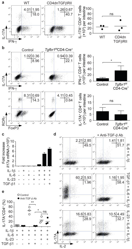
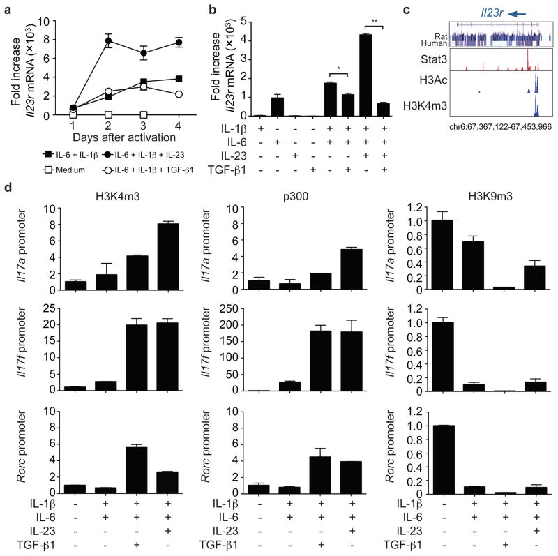
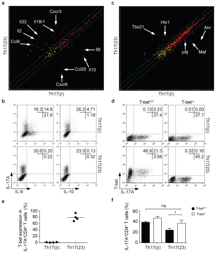
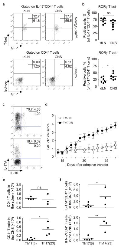
CD4(+) T-helper cells that selectively produce interleukin (IL)-17 (T(H)17), are critical for host defence and autoimmunity. Although crucial for T(H)17 cells in vivo, IL-23 has been thought to be incapable of driving initial differentiation. Rather, IL-6 and transforming growth factor (TGF)-β1 have been proposed to be the factors responsible for initiating specification. Here we show that T(H)17 differentiation can occur in the absence of TGF-β signalling. Neither IL-6 nor IL-23 alone efficiently generated T(H)17 cells; however, these cytokines in combination with IL-1β effectively induced IL-17 production in naive precursors, independently of TGF-β. Epigenetic modification of the Il17a, Il17f and Rorc promoters proceeded without TGF-β1, allowing the generation of cells that co-expressed RORγt (encoded by Rorc) and T-bet. T-bet(+)RORγt(+) T(H)17 cells are generated in vivo during experimental allergic encephalomyelitis, and adoptively transferred T(H)17 cells generated with IL-23 without TGF-β1 were pathogenic in this disease model. These data indicate an alternative mode for T(H)17 differentiation. Consistent with genetic data linking IL23R with autoimmunity, our findings re-emphasize the importance of IL-23 and therefore may have therapeutic implications.


Figures

Figure 1
Figure 1. In vivo and in vitro differentiation of Th17 cells in the absence of TGF-β signaling
a,b, Lamina propria cells were isolated from CD4dnTGFβRII and age-matched wild type (WT) mice (a) or Tgfbr1f/fCD4-Cre+ and Tgfbr1fl+CD4-Cre+ littermate controls (control) (b). Cells were stained for T cell markers and intracellular expression of IFN-γ, IL-17A, RORγt and FoxP3. Representative experiments are shown in left panels and pooled data are shown on the right (mean; error bars in b denote s.e.m., n=7). No significant differences in absolute numbers and proportions of IL-17A+CD4+ T cells were noted. *P<0.05. c–e, Naïve CD4+ T cells were isolated by cell sorting and activated in serum-free media with plate-bound anti-CD3/anti-CD28 for 4 days together with the indicated cytokines. Il17a mRNA expression was assessed by quantitative RT-PCR (c). IL-17A and IL-2 protein expression were analyzed by intracellular staining. Neutralizing anti-TGF-β antibodies prevented IL-6/IL-1β and TGF-β-dependent differentiation of Th17 cells, but not IL-23 and IL-6/IL-1β induced differentiation. Representative intracellular staining is depicted in panel d and pooled data from four individual experiments with mean values are shown in panel e. *P<0.05, **P<0.01.
Figure 2
Figure 2. IL-23 upregulates IL-23R and modifies the Il17 and Rorc loci in the absence of TGF-β
a, b, Naïve CD4+ T cells were activated in serum-free media without cytokines, with individual cytokines or cytokine combinations as indicated. Il23r expression was analyzed by quantitative RT-PCR (mRNA levels ± s.e.m.) on days 1 to 4 after activation (a) or on day 4 only (b), *P<0.01, **P<0.001. c, IL-6 and IL-23 induce Stat3 binding to Il23r, histone 3 acetylation (H3Ac) and histone 3 lysine 4 trimethylation (H3K4m3) of the Il23r locus as determined by chromatin immunoprecipitation and massive parallel sequencing. d, Naïve CD4+ cells were activated as in (a). Fixed cells were immunoprecipitated with anti-H3K4m3, anti-p300 or anti-H3K9m3 antibodies. Eluted DNA was analyzed by quantitative PCR using primers spanning the promoter regions of Il17a, Il17f and Rorc.
Figure 3
Figure 3. IL-23-induced Th17 cells express T-bet but not IL-9 and IL-10
a, Naïve CD4+ T cells were polyclonally stimulated in the presence of IL-6, IL-1β and either TGF-β (Th17(β) cells) or IL-23 (Th17(23) cells). Microarray analysis demonstrates the differential expression of genes encoding cytokines and receptors in the two subsets of Th17 cells. Mean values from two independent experiments are shown. b, Th17(β) and Th17(23) cells were polarized, expanded in IL-2, restimulated with anti-CD3/anti-CD28 antibodies (1 μg ml−1), cytokines, expanded and then analyzed for IL-17, IL-9 or IL-10 expression by intracellular staining. c, Transcription factor expression in Th17(β) and Th17(23) cells as assessed by microarray analysis. Mean values of two independent experiments are shown. d–f, Th17(23) but not Th17(β) express T-bet. Naïve CD4+ T cells were activated by IL-6, IL-1β with either IL-23 or TGF-β. IL-17 expression was not altered in T-bet−/− Th17(β) cells compared to T-bet+/+ Th17(β) cells. In contrast, loss of T-bet expression enhanced IL-17 production in Th17(23) cells. A representative experiment is depicted in d and pooled data are shown in e (n=4) and f (n=3, error bars are s.e.m., *P<0.05).
Figure 4
Figure 4. RORγt+T-bet+ Th17 cells arise during CNS inflammation and T-bet-expressing, IL-23-induced Th17 cells are more pathogenic
a, b, To induce CNS inflammation, we immunized Rorc(γt)-GfpTG or Rorc(γt)-GfpTG- control mice with MOG35–55 in CFA and CD4+ T cells isolated from the draining lymph nodes (dLN) or the CNS were analyzed by flow cytometry for IL-17, T-bet and GFP (RORγt) expression (upper panel). Lower panel shows isotype control staining for T-bet and fluorescence in transgene-negative littermates. A representative staining is depicted in a and pooled data of RORγt+ T-bet and RORγt+ T-bet+ IL-17+CD4+ T cells are shown in b (n=7, *P<0.05). c, Naïve CD4+Vβ11+CD62L+CD44 were isolated by cell sorting from TCR(2D2) transgenic mice. The cells were activated with anti-CD3/anti-CD28, IL-6, IL-1β and anti-IFN-γ neutralizing antibodies with either TGF-β1 or IL-23 with anti-TGF-β neutralizing antibodies. The resultant cells were analyzed for IL-17 and IL-10 expression by intracellular staining and flow cytometry. d, Polarized cells (1×10⁶) were adoptively transferred into Rag2−/− recipients and followed for signs of neurological disease. Data show mean ± s.e.m. of the EAE clinical score of 20 mice pooled from two independent experiments. e, f, CNS-infiltrating mononuclear cells were isolated and the total number of CD4+ T cells was determined in both groups (*P<0.01, e). The absolute numbers of CNS-infiltrating IL-17+ or IFN-γ+ CD4+ T cells of each group were assessed by intracellular cytokine staining (*P<0.05, **P<0.01, f).

Comment in

References

    1. Miossec P, Korn T, Kuchroo VK. Interleukin-17 and type 17 helper T cells. N Engl J Med. 2009;361:888–898. - PubMed
    1. Weaver CT, Hatton RD, Mangan PR, Harrington LE. IL-17 family cytokines and the expanding diversity of effector T cell lineages. Annu Rev Immunol. 2007;25:821–852. - PubMed
    1. Stockinger B, Veldhoen M. Differentiation and function of Th17 T cells. Curr Opin Immunol. 2007;19:281–286. - PubMed
    1. Zhou L, Chong MM, Littman DR. Plasticity of CD4+ T cell lineage differentiation. Immunity. 2009;30:646–655. - PubMed
    1. Cua DJ, et al. Interleukin-23 rather than interleukin-12 is the critical cytokine for autoimmune inflammation of the brain. Nature. 2003;421:744–748. - PubMed

Publication types

MeSH terms

Associated data