. 2011 May 13;30(13):2569-81.
doi: 10.1038/emboj.2011.151.

AP-2γ regulates oestrogen receptor-mediated long-range chromatin interaction and gene transcription

Affiliations

AP-2γ regulates oestrogen receptor-mediated long-range chromatin interaction and gene transcription

Si Kee Tan et al. EMBO J. .

Erratum in

  • EMBO J. 2011;30(13):2750. Sung, Win King [corrected to Sung, Wing Kin]

Abstract

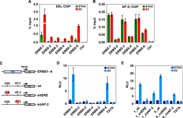
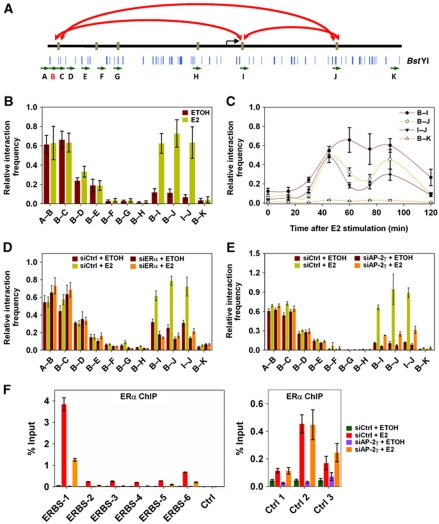
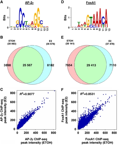
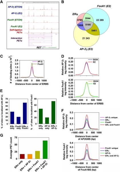
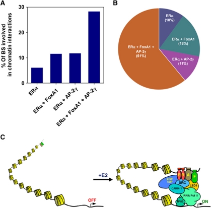
Oestrogen receptor α (ERα) is key player in the progression of breast cancer. Recently, the cistrome and interactome of ERα were mapped in breast cancer cells, revealing the importance of spatial organization in oestrogen-mediated transcription. However, the underlying mechanism of this process is unclear. Here, we show that ERα binding sites (ERBS) identified from the Chromatin Interaction Analysis-Paired End DiTag of ERα are enriched for AP-2 motifs. We demonstrate the transcription factor, AP-2γ, which has been implicated in breast cancer oncogenesis, binds to ERBS in a ligand-independent manner. Furthermore, perturbation of AP-2γ expression impaired ERα DNA binding, long-range chromatin interactions, and gene transcription. In genome-wide analyses, we show that a large number of AP-2γ and ERα binding events converge together across the genome. The majority of these shared regions are also occupied by the pioneer factor, FoxA1. Molecular studies indicate there is functional interplay between AP-2γ and FoxA1. Finally, we show that most ERBS associated with long-range chromatin interactions are colocalized with AP-2γ and FoxA1. Together, our results suggest AP-2γ is a novel collaborative factor in ERα-mediated transcription.


Conflict of interest statement

The authors declare that they have no conflict of interest.

Figures

Figure 1
Figure 1
AP-2γ is required for transcription of oestrogen-regulated genes. (A) Screenshot of ERα ChIA-PET analysis showing ERα binding and long-range chromatin interactions at the RET gene locus. ERBS are represented as density histogram (red) and long-range chromatin interactions are represented as intra-chromosomal interaction PETs (magenta). RET-associated ERBS are denoted by numbers (blue). (B) MCF-7 cells were stimulated with or without E2 for 0, 3, 6, 12, and 24 h and then analysed by reverse transcription and real-time RT–PCR for the level of RET 9 and 51 mRNA expressions. (C) MCF-7 cells were transfected with control, ERα or AP-2γ siRNA, stimulated with or without E2 for 12 h and then analysed for RET 9 and 51 mRNA levels. (D) Gene expression profiling was performed on MCF-7 cells that were transfected with control or AP-2γ siRNA and stimulated with or without E2 for 12 h. The heatmap represents all E2-regulated genes and fold change in expression is indicated below. E2-upregulated genes and E2-downregulated genes that are no longer activated and repressed due to AP-2γ knockdown are marked by the red and blue asterisks, respectively. All results represent the average of three independent experiments ±s.e.m.
Figure 2
Figure 2
Ligand-independent recruitment of AP-2γ at RET ERBS. ChIP assays were performed in MCF-7 cells treated with or without E2 for 45 min using antibodies against (A) ERα and (B) AP-2γ. Binding was assessed by real-time RT–PCR at RET-associated ERBS as described in Figure 1A. (C) Schematic diagram showing the reporter constructs that were generated and used in transient transfection analysis. (D) MCF-7 cells were transfected with reporter constructs and treated with or without E2 for 24 h. Luciferase assays were performed using a dual-luciferase system with Renilla as an internal control. The six RET-associated ERBS, ERBS-1–6, were each cloned into pGL4-TATA and assessed in transient transfection analysis. (E) ERE and AP-2 motifs predicted in ERBS-1 and -6 were mutated and compared with their equivalent wild-type versions in transient transfection analysis. All results represent the average of three independent experiments ±s.e.m.
Figure 3
Figure 3
AP-2γ is required for efficient ERα binding and long-range chromatin interactions. (A) Schematic diagram showing the location of primers (green arrows) and BstYI restriction enzyme cutting sites (blue lines) at the regulatory region of the RET gene. Primer B (red) was used as the main ‘anchor’ region for the 3C assay. Long-range chromatin interactions detected by the 3C assay are indicated by red arrows. (B) 3C assay was performed on MCF-7 cells treated with or without E2 for 45 min. Interactions were detected by real-time PCR using primers indicated in (A). (C) MCF-7 cells were exposed to E2 for 0, 15, 30, 45, 60, 75, 90, and 120 min and then examined by 3C analysis. MCF-7 cells were transfected with (D) ERα, (E) AP-2γ, and control siRNA, treated with or without E2 for 45 min, and then subjected to 3C analysis. (F) ChIP assay using ERα antibody was performed on MCF-7 cells transfected with control or AP-2γ siRNA and treated with or without E2 for 45 min. ERα binding was assessed at the RET-associated ERBS and at control ERBS that do not coincide with AP-2γ binding (right panel). All results represent the average of three independent experiments ±s.e.m.
Figure 4
Figure 4
Global analysis of AP-2γ and FoxA1 binding events in MCF-7 cells. (A, D) De novo identification of the AP-2 and FoxA1 binding motif with the top 500 AP2GBS and FoxA1BS (±50 bp of sequence from the ChIP-seq peak) using MEME. (B, E) Comparison of AP2GBS and FoxA1BS overlap (with a window size of ±250 bp) under vehicle or E2 conditions. (C, F) Scatter plots representing the correlation of peak intensities of AP-2γ and FoxA1 before and after E2 stimulation.
Figure 5
Figure 5
AP-2γ, FoxA1, and ERα are colocalized at a large fraction of ERBS. (A) Screenshot showing tracks from the ChIA-PET of ERα (magenta) and ChIP-seq profiles of AP-2γ (blue) and FoxA1 (green) at the RET gene locus. (B) Venn diagram showing overlap of ChIA-PET ERBS, AP2GBS, and FoxA1BS within ±250 bp of each respective peak binding location. (C) Frequency of AP-2γ and FoxA1 peak distribution with respect to the centre of ERBS (50 bp bin size). (D) Distribution of the average AP-2γ and FoxA1 ChIP-seq tag intensity before and after E2 stimulation was examined with respect to the centre of ERBS (±1 kb with 100 bp bin size). ChIA-PET ERBS, AP2GBS, and FoxA1BS were clustered and different overlapping regions were analysed for (E) the frequency of AP2GBS and FoxA1BS occurrence, (F) the average tag intensity distribution of AP-2γ and FoxA1 (±1 kb with 100 bp bin size), and (G) the average PET count from the ERα ChIA-PET data set.
Figure 6
Figure 6
Mutual requirement of AP-2γ and FoxA1 recruitment at ERBS. (A) ChIP for FoxA1 was performed on MCF-7 cells treated with or without E2 for 45 min. (B) ChIP for FoxA1 was performed on MCF-7 cells transfected with control or AP-2γ siRNA. (C) ChIP for AP-2γ was performed on MCF-7 cells transfected with control or FoxA1 siRNA. FoxA1 and AP-2γ binding were examined at RET-associated ERBS and at control ERBS that FoxA1 and AP-2γ do not colocalize. All results represent the average of three independent experiments ±s.e.m.
Figure 7
Figure 7
ERα collaborates with AP-2γ and FoxA1 to promote long-range chromatin interactions across the genome. (A) Percentages of different overlapping ERBS regions that are involved in chromatin interactions. (B) Pie chart showing the proportion of interacting ERBS that are unique or colocalized with AP-2γ and/or FoxA1 binding. (C) Schematic diagram illustrating how AP-2γ and FoxA1 facilitates and coordinate ERα transcriptional activity. AP-2γ and FoxA1 are pre-recruited to the ERBS where they both work cooperatively to promote ERα binding and subsequent chromatin looping, finally stimulating transcription. AP-2γ and FoxA1 are denoted by ‘A’ and ‘F’, respectively.

References

    1. Ali S, Coombes RC (2000) Estrogen receptor alpha in human breast cancer: occurrence and significance. J Mammary Gland Biol Neoplasia 5: 271–281 - PubMed
    1. Arighi E, Borrello MG, Sariola H (2005) RET tyrosine kinase signaling in development and cancer. Cytokine Growth Factor Rev 16: 441–467 - PubMed
    1. Boulay A, Breuleux M, Stephan C, Fux C, Brisken C, Fiche M, Wartmann M, Stumm M, Lane HA, Hynes NE (2008) The Ret receptor tyrosine kinase pathway functionally interacts with the ERalpha pathway in breast cancer. Cancer Res 68: 3743–3751 - PubMed
    1. Carroll JS, Liu XS, Brodsky AS, Li W, Meyer CA, Szary AJ, Eeckhoute J, Shao W, Hestermann EV, Geistlinger TR, Fox EA, Silver PA, Brown M (2005) Chromosome-wide mapping of estrogen receptor binding reveals long-range regulation requiring the forkhead protein FoxA1. Cell 122: 33–43 - PubMed
    1. Carroll JS, Meyer CA, Song J, Li W, Geistlinger TR, Eeckhoute J, Brodsky AS, Keeton EK, Fertuck KC, Hall GF, Wang Q, Bekiranov S, Sementchenko V, Fox EA, Silver PA, Gingeras TR, Liu XS, Brown M (2006) Genome-wide analysis of estrogen receptor binding sites. Nat Genet 38: 1289–1297 - PubMed

Publication types

MeSH terms

Associated data