. 2011 Apr 29;145(3):435-46.
doi: 10.1016/j.cell.2011.03.044.

Nucleotide deficiency promotes genomic instability in early stages of cancer development

Affiliations

Nucleotide deficiency promotes genomic instability in early stages of cancer development

Assaf C Bester et al. Cell. .

Abstract

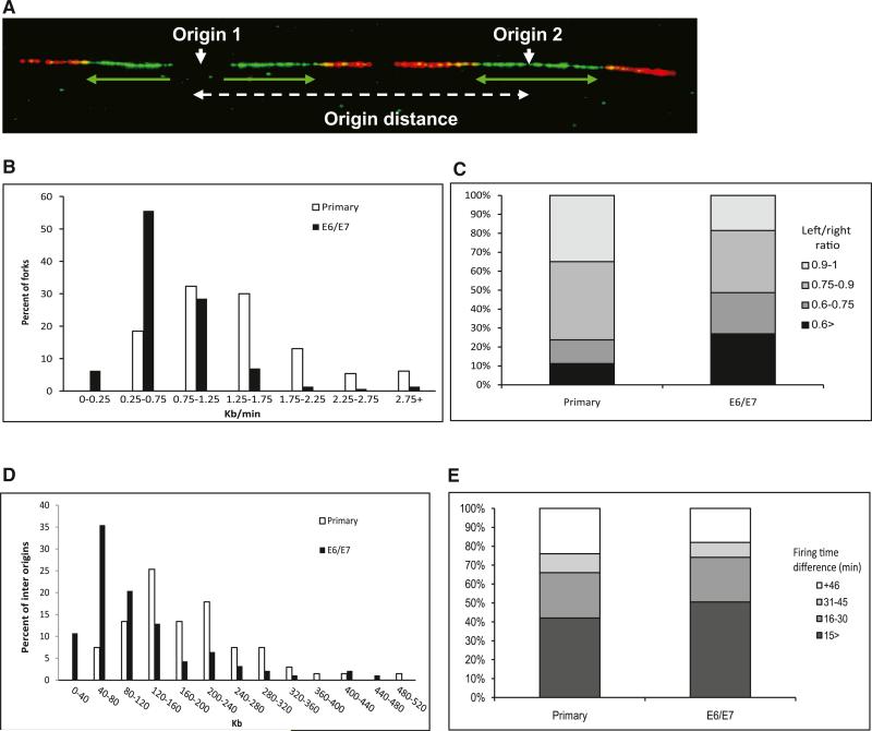
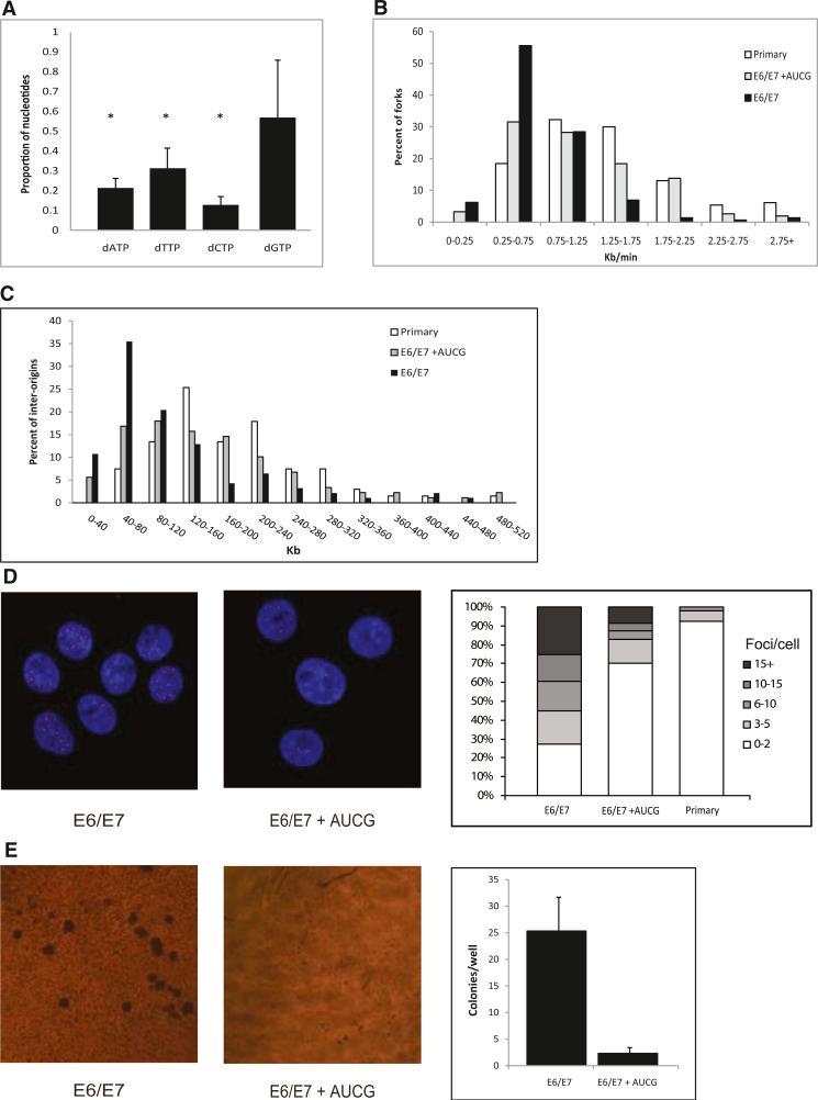
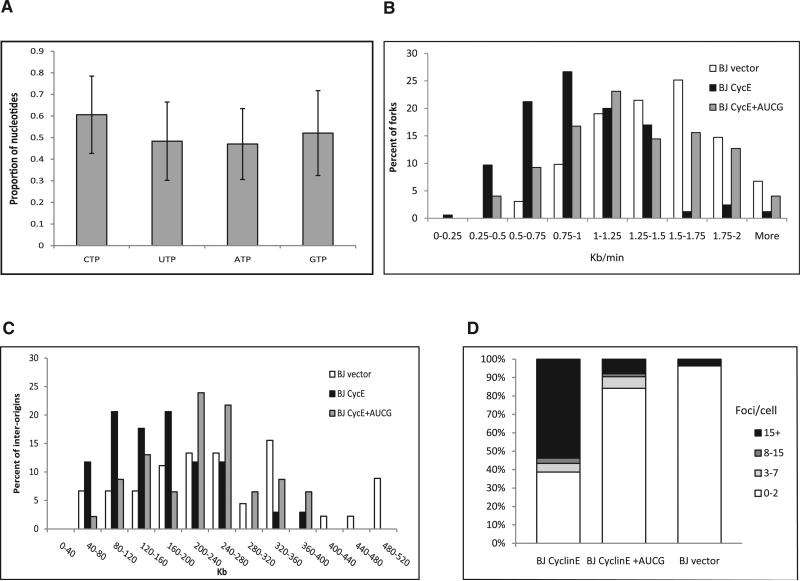
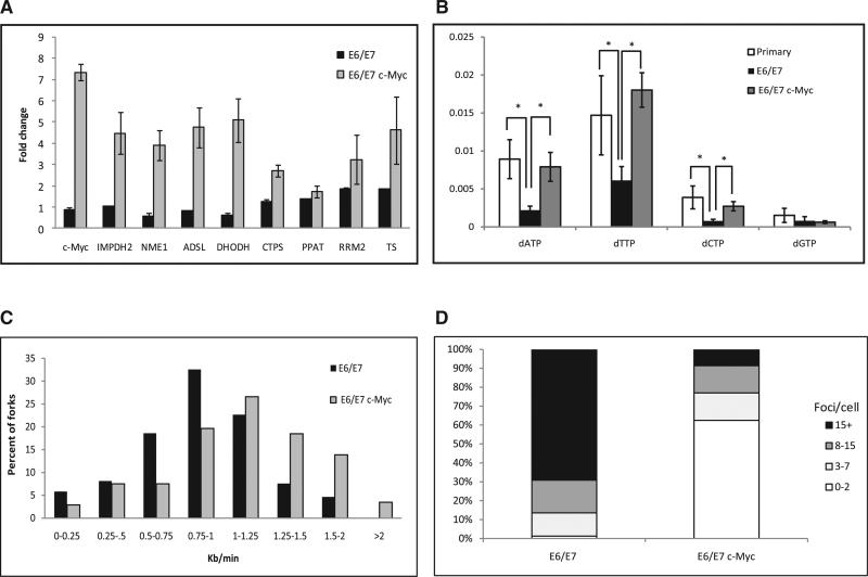
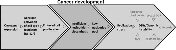
Chromosomal instability in early cancer stages is caused by stress on DNA replication. The molecular basis for replication perturbation in this context is currently unknown. We studied the replication dynamics in cells in which a regulator of S phase entry and cell proliferation, the Rb-E2F pathway, is aberrantly activated. Aberrant activation of this pathway by HPV-16 E6/E7 or cyclin E oncogenes significantly decreased the cellular nucleotide levels in the newly transformed cells. Exogenously supplied nucleosides rescued the replication stress and DNA damage and dramatically decreased oncogene-induced transformation. Increased transcription of nucleotide biosynthesis genes, mediated by expressing the transcription factor c-myc, increased the nucleotide pool and also rescued the replication-induced DNA damage. Our results suggest a model for early oncogenesis in which uncoordinated activation of factors regulating cell proliferation leads to insufficient nucleotides that fail to support normal replication and genome stability.


Figures

Figure 1
Figure 1. Replication Dynamics in Primary and HPV-16 E6/E7-Expressing Keratinocytes
(A) Example of a single combed DNA molecule labeled with IdU (green) and CldU (red), showing replication from two adjacent origins. Green arrows, orientation and length of fork progression. (B) Fork rate (Kb/min) distribution. White bars, primary keratinocytes (n = 130); black bars, keratinocytes expressing E6/E7 (n = 144). (C) Progression ratio between left and right forks emerging from the same origin. Primary keratinocytes (n = 156); keratinocyte-expressing E6/E7 (n = 194). (D) Origin density distribution. White bars, primary keratinocytes (n = 67); black bars, keratinocytes expressing E6/E7 (n = 93). (E) Differences in firing time between adjacent origins. The groups represent different relative firing time in minutes. Primary keratinocytes (n = 50); keratinocytes expressing E6/E7 (n = 89). See also Figure S1, Figure S2, Table S1, and Table S2.
Figure 2
Figure 2. The Effect of an Exogenous Supply of Nucleosides on the Replication Dynamics, DNA Damage, and Transformation in HPV-16 E6/E7-Expressing Cells
(A) The nucleotide levels in keratinocyte cells expressing E6/E7 normalized to the level in primary cells of the same donor. *p < 0.05. The levels are expressed as mean fold change ± SEM (n = 3). (B) Fork rate (Kb/min) distribution. White bars, primary keratinocytes (n = 130); black bars, keratinocytes expressing E6/E7 (n = 150); gray bars, keratinocytes expressing E6/E7 grown for 48 hr with exogenous supply of nucleosides (n = 155). (C) Origin density (Kb) distribution. White bars, primary keratinocytes (n = 67); black bars, keratinocytes expressing E6/E7 (n = 93); gray bars, keratinocytes expressing E6/E7 grown for 48 hr with exogenous supply of nucleosides (n = 89). (D) (Left) Examples of nuclei with γH2AX foci following expression of E6/E7 grown in a normal medium (E6/E7) and in a medium supplied with exogenous nucleosides (E6/E7+AUCG). The nuclei were stained with DAPI. (Right) Percent of nuclei with the indicated number of γH2AX foci. Primary keratinocyte cells (n = 51); keratinocytes expressing E6/E7 (n = 52); keratinocytes expressing E6/E7 supplied with exogenous nucleosides (n = 70). (E) (Left) Examples of anchorage-independent growth in soft agar. (Right) Mean number of colonies per a soft agar plate. Cells expressing E6/E7; cells expressing E6/E7 supplied with exogenous nucleosides. The number of colonies per plate is expressed as mean ± SEM (n = 3). See also Figure S3 and Figure S4, and Table S3.
Figure 3
Figure 3. The Effect of an Exogenous Supply of Nucleosides on the Replication Dynamics and DNA Damage of BJ Cells Expressing Cyclin E
(A) rNTP pool following cyclin E expression in BJ cells. The levels are expressed as mean fold change ± SEM (n = 4). (B) Fork rate distribution (Kb/min). White bars, BJ cells (n = 163); black bars, BJ expressing cyclin E (n = 165); gray bars, BJ expressing cyclin E grown for 48 hr with exogenous supply of nucleosides (n = 173). (C) Origin density distribution (Kb). White bars, BJ cells (n = 45); black bars, BJ expressing cyclin E (n = 34); gray bars, BJ expressing cyclin E grown for 48 hr with exogenous supply of nucleosides (n = 46). (D) Percent of nuclei with the indicated number of γH2AX foci. BJ cells (n = 57); BJ expressing cyclin E (n = 110); BJ expressing cyclin E grown for 48 hr with exogenous supply of nucleosides (n = 64). See also Figure S5 and Table S3.
Figure 4
Figure 4. The Effect of c-myc Expression on the Expression of the Nucleotide Biosynthesis Genes, Nucleotide Pools, Replication Dynamics, and DNA Damage
(A) Fold change in the expression level of c-Myc targets from the nucleotide biosynthesis pathways. Black bars, expression of E6/E7 normalized to the level in primary keratinocytes; gray bars, expression of c-myc in E6/E7-expressing keratinocytes normalized to the level in cells expressing only E6/E7. The levels are expressed as mean fold change ± SEM (n = 3). (B) Nucleotide levels in primary keratinocytes, keratinocytes expressing E6/E7, and keratinocytes coexpressing E6/E7 and c-myc. *p < 0.05. The levels are expressed as mean fold change ± SEM (n > 3). (C) Fork rate distribution (Kb/min). Black bars, keratinocytes expressing E6/E7 (n = 173); gray bars, keratinocytes expressing E6/E7 with enhanced nucleotide biosynthesis by c-Myc (n = 175). (D) Percent of nuclei with the indicated number of γH2AX foci in cells expressing E6/E7 (n = 80) and in keratinocytes expressing E6/E7 with enhanced nucleotide biosynthesis by c-Myc (n = 103). See also Figure S6.
Figure 5
Figure 5. A Model for the Events Leading to Genomic Instability in Early Stages of Cancer Development
Oncogene expression forces cell proliferation by aberrant activation of cell-cycle regulators (Rb-E2F). Insufficient activation of the nucleotide biosynthesis pathways results in a low-nucleotide pool that fails to support normal DNA replication. This leads to replication stress and promotes genomic instability during early stages of cancer development. Additional factors contribute to genomic instability in different stages of tumorigenesis, such as reactive oxygen species (ROS), telomere loss, hypoxia, abrogated mitotic checkpoints, and loss of the DNA damage response (DDR).

Comment in

References

    1. Akli S, Keyomarsi K. Cyclin E and its low molecular weight forms in human cancer and as targets for cancer therapy. Cancer Biol. Ther. 2003;2(4, Suppl 1):S38–S47. - PubMed
    1. Anglana M, Apiou F, Bensimon A, Debatisse M. Dynamics of DNA replication in mammalian somatic cells: nucleotide pool modulates origin choice and interorigin spacing. Cell. 2003;114:385–394. - PubMed
    1. Bartkova J, Horejsí Z, Koed K, Krämer A, Tort F, Zieger K, Guldberg P, Sehested M, Nesland JM, Lukas C, et al. DNA damage response as a candidate anti-cancerbarrierinearly humantumorigenesis. Nature. 2005;434:864–870. - PubMed
    1. Bartkova J, Rezaei N, Liontos M, Karakaidos P, Kletsas D, Issaeva N, Vassiliou LV, Kolettas E, Niforou K, Zoumpourlis VC, et al. Onco-gene-induced senescence is part of the tumorigenesis barrier imposed by DNA damage checkpoints. Nature. 2006;444:633–637. - PubMed
    1. Bartkova J, Hamerlik P, Stockhausen MT, Ehrmann J, Hlobilkova A, Laursen H, Kalita O, Kolar Z, Poulsen HS, Broholm H, et al. Replication stress and oxidative damage contribute to aberrant constitutive activation of DNA damage signalling in human gliomas. Oncogene. 2010;29:5095–5102. - PubMed

Publication types

Associated data