. 2011 Jul 22;146(2):318-31.
doi: 10.1016/j.cell.2011.06.019. Epub 2011 Jul 14.

Generation of isogenic pluripotent stem cells differing exclusively at two early onset Parkinson point mutations

Affiliations

Generation of isogenic pluripotent stem cells differing exclusively at two early onset Parkinson point mutations

Frank Soldner et al. Cell. .

Erratum in

  • Cell. 2011 Aug 19;146(4):659

Abstract

Patient-specific induced pluripotent stem cells (iPSCs) derived from somatic cells provide a unique tool for the study of human disease, as well as a promising source for cell replacement therapies. One crucial limitation has been the inability to perform experiments under genetically defined conditions. This is particularly relevant for late age onset disorders in which in vitro phenotypes are predicted to be subtle and susceptible to significant effects of genetic background variations. By combining zinc finger nuclease (ZFN)-mediated genome editing and iPSC technology, we provide a generally applicable solution to this problem, generating sets of isogenic disease and control human pluripotent stem cells that differ exclusively at either of two susceptibility variants for Parkinson's disease by modifying the underlying point mutations in the α-synuclein gene. The robust capability to genetically correct disease-causing point mutations in patient-derived hiPSCs represents significant progress for basic biomedical research and an advance toward hiPSC-based cell replacement therapies.


Figures

Figure 1
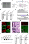
Figure 1. ZFN-mediated insertion of the A53T (G209) α-synuclein mutation in hESCs using a drug selection based targeting strategy
(A) Screening of ZFNs-directed against nucleotide 209 of exon 3 of human α-synuclein. ZFNs mediated disruption of target locus was measured by the Surveyor/Cel-1 assay. Red arrow indicates expected Cel-1 digest product. The frequency of gene disruption of each ZFN pair is indicated below each lane (GFP indicates GFP transfected negative control). All ZFNs were linked to wild-type FokI, except pair SNCA-L1/R3, which was linked to an obligate heterodimer form of the Fok1 endonuclease (ELD-KKR) (B) Schematic overview depicting the genomic α-synuclein locus (SNCA) and the targeting strategy showing exons (blue boxes), restriction sites and location of external and internal southern blot probes (red bars). Enlarged sequence indicates ZFNs induced cut site at base 209 in exon 3 of α-synuclein (red base pair) and insertion site of loxP-site-flanked pGK-puro selection cassette (red box). Shown below is a schematic of the donor plasmid design for either positive selection (Syn-A53T-loxP-pGK-puro-loxP) or positive-negative selection (Syn-A53T-loxP-pGK-puro-loxP-HSV-TK-DT-A) and targeted genomic locus before and after Cre-excision of the selection cassette. Donor plasmids comprise ~600 bp homology on each side of the ZFN cut. pGK-promoter, phosphoglycerol kinase promoter; puro, puromycin resistance gene; pGKpolyA, polyadenylation sequence; HSV-TK, herpes simplex virus thymide kinase; pGK-DT-A-pA, diphteria toxin A-chain. (C) Southern blot analysis of hESC line WIBR3 targeted with donor plasmid Syn-A53T-loxP-pGK-puro-loxP before (WIBR3-SNCAA53T/WT-1) and after (WIBR3-SNCAA53T/WT-1C) Cre-mediated excision of the selection cassette. Genomic DNA was digested with indicated enzymes and hybridized with the external 3′ and 5′ probe and internal 3′ probe. Fragment sizes for each digest are indicated. (D) Sequencing of genomic α-synuclein locus in hESC line WIBR3-SNCAA53T/WT-1C showing either wild-type (G209, blue base) or targeted mutant (A209, red base) sequence. Targeted allele contains the remaining loxP site after Cre-mediated excision of selection cassette. (E) Immunofluorescence staining of WIBR3-SNCAA53T/WT-1C for the pluripotency markers OCT4, NANOG, SOX2, Tra-1-60, Tra-1-81 and SSEA4. (F) Hematoxylin and eosin staining of teratoma sections generated from WIBR3-SNCAA53T/WT-1C cells. (G) Immunofluorescence staining of neuronal cultures derived from WIBR3-SNCAA53T/WT-1C cells 10 days after induction of differentiation for neuron specific class III β-tubulin (TUJ1; green) and the dopaminergic neuron specific marker tyrosine hydroxylase (TH; red). (H) Mutation analysis RT-PCR of α-synuclein transcript (+/−Tsp45I restriction digest) in indicated cell lines. (A53T mutation creates additional Tsp45I restriction site). Expected fragment size after Tsp45I digest: wild-type transcript, 249/218/24/9 bp; A53T transcript 249/185/33/24/9 bp (A53T and wild-type (wt) derived restriction fragments are indicated by red arrows). (I) Southern blot analysis of WIBR3 cells targeted with donor plasmid Syn-A53T-loxP-pGK-puro-loxP-HSV-TK-DT-A after positive-negative selection. Genomic DNA was digested with indicated enzymes and hybridized with the external 5′ and 3′ probe and internal 3′ probe. Fragment sizes for each digest are indicated (WIBR3-SNCAA53T/WT-2 line shows correct targeting of one allele; WIBR3-SNCAA53T/A53T line shows correct targeting of both alleles).
Figure 2
Figure 2. ZFN-mediated insertion of the A53T α-synuclein mutation in hESC line BGO1 without drug-selection
(A) Schematic overview depicting the genomic α-synuclein (SNCA) locus and the targeting strategy showing exons (blue boxes), restriction sites and location of internal southern blot probe (red bars). Enlarged sequence indicates ZFNs induced cut site at base 209 in exon 3 of α-synuclein (red base pair). Shown below is a schematic of donor plasmid design and targeted genomic locus for either insertion (Donor-A53T) or correction (wild-type Donor) of A53T (G209A) α-synuclein mutation. Donor plasmids contain ~1 kb homology to the targeting site. (B) Southern blot screening (3′ internal probe) of BGO1 cells for targeted integration of A53T (G209A) mutation (Donor-A53T). Genomic DNA was digested using an A53T (G209A)-allele-specific Tsp45I restriction digest (A53T mutation creates additional Tsp45I restriction site). Expected fragment sizes wild-type allele, 2.96 kb; A53T allele, 1.55 kb. 3′ internal southern blot probe excludes additional non-homologous integration of the donor vector. Red asterisk indicates clone with additional Tsp45I restriction site indicative of insertion of A53T mutation. (C) PCR mutation analysis of α-synuclein locus (+/−Tsp45I restriction digest) in patient derived hiPSCs (WIBR-iPS-SNCAA53T) and targeted hESCs (BGO1-SNCAA53T/WT represents clone marked by red asterisk in (B)). Fragment sizes (+Tsp45I) wild-type allele, 219 bp; A53T allele, 131/88bp. (D) Sequencing of genomic α-synuclein locus in targeted hESC clone BGO1-SNCAA53T/WT displaying either wild-type allele (G209 in 7 out of 12 sequences) or targeted mutant A53T allele (A209 in 5 out of 12 sequences). (E) Immunofluorescence staining of BGO1-SNCAA53T/WT for the pluripotency markers OCT4, NANOG, SOX2, Tra-1-60, Tra-1-81 and SSEA4. (F) Hematoxylin and eosin staining of teratoma sections generated from WIBR3-SNCAA53T/WT-1C cells. (G) Immunofluorescence staining of neuronal cultures derived from WIBR3-SNCAA53T/WT-1C cells 10 days after induction of differentiation for neuron specific class III β-tubulin (TUJ1; green) and the dopaminergic neuron specific marker tyrosine hydroxylase (TH; red).
Figure 3
Figure 3. ZFN-mediated insertion of the E46K α-synuclein mutation in hESCs using ssODNs
(A) Schematic overview depicting the genomic α-synuclein (SNCA) locus and the targeting strategy showing exon 3 (blue boxe), restriction sites and location of southern blot probe (red bar). Enlarged sequence indicates ZFNs induced cut site at base 209 in exon 3 of α-synuclein and the site of the E46K (G188A) mutation (red base pair). Shown below is a schematic of the 114bp ssODN and the targeted genomic locus after insertion of E46K (G188A) α-synuclein mutation. (B) PCR mutation analysis of α-synuclein locus (+/−StyI restriction digest) in hESC lines WIBR3 and BGO1 and targeted E46K hESC lines (WIBR3-SNCAE46K and BGO1-SNCAE46K ). Fragment sizes (+ StyI) wild-type allele, 153/66bp, E46K mutated allele 219 bp. (C) Southern blot analysis (3′-probe) of WIBR3 and BGO1 and targeted E46K hESC lines (WIBR3-SNCAE46K and BGO1-SNCAE46K) for targeted integration of A53T (G209A) mutation. Genomic DNA was digested using an E46K (G188A)-allele-specific StyI restriction digest (E46K mutation disrupts wild-type allele specific StyI restriction site). Expected fragment sizes wild-type allele, 0.46 kb; E46K mutated allele, 2.96 kb). (D) Sequencing of genomic α-synuclein locus in targeted hESC clone WIBR3-SNCAE46K displaying either wild-type allele (G188 in 10 out of 16 sequences) or targeted mutant E46K allele (A188 in 6 out of 16 sequences). (E) Sequencing of genomic α-synuclein locus in targeted hESC clone BGO1-SNCAE46K displaying either wild-type allele (G188 in 7 out of 16 sequences) or targeted mutant E46K allele (A188 in 9 out of 16 sequences). (F) Immunofluorescence staining of WIBR3-SNCAE46K for the pluripotency markers OCT4, NANOG, SOX2, Tra-1-60, Tra-1-81 and SSEA4. (G) Immunofluorescence staining of BGO1-SNCAE46K for the pluripotency markers OCT4, NANOG, SOX2, Tra-1-60, Tra-1-81 and SSEA4.
Figure 4
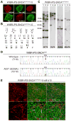
Figure 4. Derivation of disease-specific hiPSCs from PD patient with A53T (G209A) α-synuclein mutation
(A) Immunofluorescence staining of PD patient-derived hiPSC line WIBR-iPS-SNCAA53T(2lox)-5 for the pluripotency markers OCT4, NANOG, SOX2, Tra-1-60, Tra-1-81 and SSEA4. (B) Cytogenetic analysis of patient-derived hiPSC line WIBR-iPS-SNCAA53T(2lox)-5 showing a normal karyotype. (C) Southern blot analysis of WIBR-iPS-SNCAA53T(2lox) clones. Genomic DNA was digested with XbaI and probed for proviral integrations with 32P-labeled DNA probes against hOCT4, hKLF4, hSOX2 and M2rtTA. Southen blot analysis confirms independent clones based on individual proviral integration patterns. (D) Sequencing of genomic α-synuclein locus in PD patient-derived hiPSCs line WIBR-iPS-SNCAA53T(2lox)-5 showing both wild-type allele (G209 in 7 out of 19 sequences) and mutant A53T allele (A209 in 12 out of 19 sequences). (E) Immunofluorescence staining of neuronal cultures derived from hiPSC line WIBR-iPS-SNCAA53T(2lox)-5 10 days after induction of differentiation for neuron specific class III β-tubulin (TUJ1; green) and the dopaminergic neuron specific marker tyrosine hydroxylase (TH; red).
Figure 5
Figure 5. ZFN mediated correction of A53T (G209A) mutation in PD patient derived hiPSCs
(A) Southern blot screening (3′ internal probe) of patient-derived hiPSCs (WIBR-iPS-SNCAA53T) for correction of A53T (G209A) mutation (wild-type Donor). Genomic DNA was digested using an A53T (G209A)-allele-specific Tsp45I restriction digest (A53T mutation creates additional Tsp45I restriction site). 3′ internal southern blot probe excludes additional non-homologous integration of the donor vector. Fragment sizes: wild-type allele, 2.96 kb; A53T allele, 1.55 kb. Red asterisk indicates clone with loss of Tsp45I restriction site indicative of loss of A53T mutation. (B) PCR mutation analysis of genomic α-synuclein locus (+/−Tsp45I restriction digest) in hESCs (WIBR3) and patient-derived hiPSCs before (WIBR-iPS-SNCAA53T) and after correction (WIBR-iPS-SNCAA53T-Corr-1 and 5, representing two sub-clones from clone marked by red asterisk in (A)). Fragment sizes (+Tsp45I): wild-type allele, 219 bp; A53T mutant allele, 131/88bp. (C) Sequencing of genomic α-synuclein locus in PD patient-derived hiPSCs after ZFN mediated correction. Corrected cell lines (WIBR-iPS-SNCAA53T-Corr-1 and 5) show only wild-type allele (G209 in 35 out of 35 sequences). (D) Mutation analysis RT-PCR of α-synuclein transcript (+/−Tsp45I restriction digest) in indicated cell lines. (A53T mutation creates additional Tsp45I restriction site). Expected fragment size after Tsp45I digest: wild-type transcript, 249/218/24/9 bp; A53T transcript 249/185/33/24/9 bp (A53T and wild-type (wt) derived restriction fragments are indicated by red arrows). (E) Cytogenetic analysis of corrected PD patient-derived hiPSC line WIBR-iPS-SNCAA53T-Corr-5 showing a normal karyotype. (F) Immunofluorescence staining of WIBR-iPS-SNCAA53T-Corr-5 line for the pluripotency markers OCT4, NANOG, SOX2, Tra-1-60, Tra-1-81 and SSEA4. (G) Hematoxylin and eosin staining of teratoma sections generated from WIBR-iPS-SNCAA53T-Corr-5 cells. (H) Immunofluorescence staining of neuronal cultures derived from WIBR3-SNCAA53T/WT-1C cells 10 days after induction of differentiation for neuron specific class III β-tubulin (TUJ1; green) and the dopaminergic neuron specific marker tyrosine hydroxylase (TH; red).

Comment in

References

    1. Bock C, Kiskinis E, Verstappen G, Gu H, Boulting G, Smith ZD, Ziller M, Croft GF, Amoroso MW, Oakley DH, et al. Reference Maps of human ES and iPS cell variation enable high-throughput characterization of pluripotent cell lines. Cell. 2011;144:439–452. - PMC - PubMed
    1. Boulting GL, Kiskinis E, Croft GF, Amoroso MW, Oakley DH, Wainger BJ, Williams DJ, Kahler DJ, Yamaki M, Davidow L, et al. A functionally characterized test set of human induced pluripotent stem cells. Nat Biotechnol. 2011;29:279–286. - PMC - PubMed
    1. Brennand KJ, Simone A, Jou J, Gelboin-Burkhart C, Tran N, Sangar S, Li Y, Mu Y, Chen G, Yu D, et al. Modelling schizophrenia using human induced pluripotent stem cells. Nature 2011 - PMC - PubMed
    1. Capecchi MR. Altering the genome by homologous recombination. Science. 1989;244:1288–1292. - PubMed
    1. Daley GQ, Scadden DT. Prospects for stem cell-based therapy. Cell. 2008;132:544–548. - PubMed

Publication types

Associated data