. 2012 Nov 1;371(1):1-12.
doi: 10.1016/j.ydbio.2012.06.005. Epub 2012 Jul 2.

E-cadherin is required for intestinal morphogenesis in the mouse

Affiliations

E-cadherin is required for intestinal morphogenesis in the mouse

Benjamin J Bondow et al. Dev Biol. .

Abstract

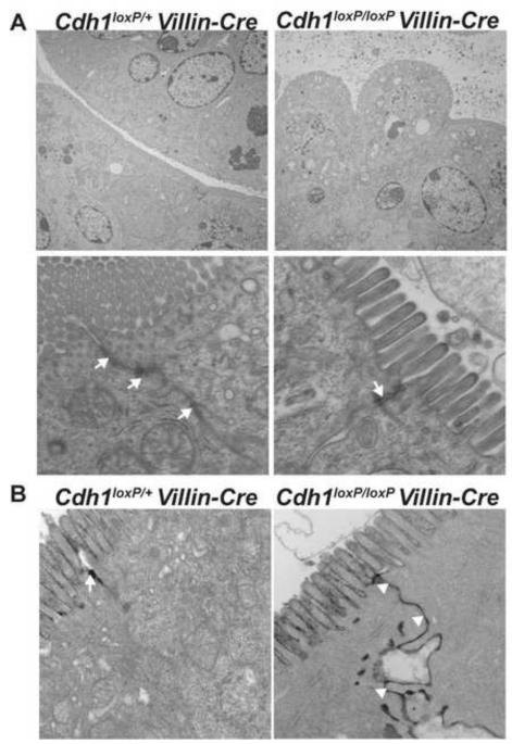
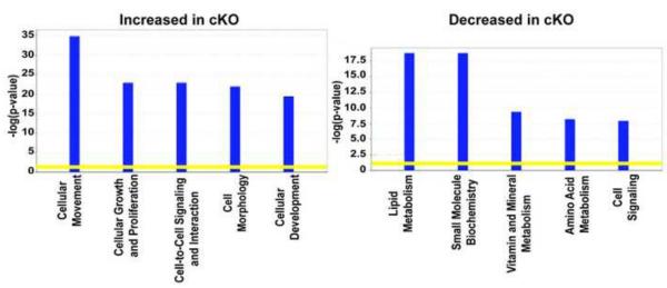
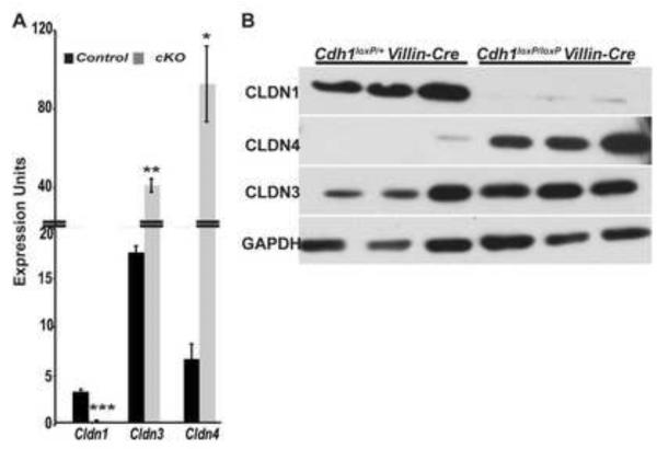
E-cadherin, the primary epithelial adherens junction protein, has been implicated as playing a critical role in nucleating formation of adherens junctions, tight junctions, and desmosomes. In addition to its role in maintaining structural tissue integrity, E-cadherin has also been suggested as an important modulator of cell signaling via interactions with its cytoplasmic binding partners, catenins, as well as with growth factor receptors. Therefore, we proposed that loss of E-cadherin from the developing mouse intestinal epithelium would disrupt intestinal epithelial morphogenesis and function. To test this hypothesis, we used a conditional knockout approach to eliminate E-cadherin specifically in the intestinal epithelium during embryonic development. We found that E-cadherin conditional knockout mice failed to survive, dying within the first 24 hours of birth. Examination of intestinal architecture at E18.5 demonstrated severe disruption to intestinal morphogenesis in animals lacking E-cadherin in the epithelium of the small intestine. We observed changes in epithelial cell shape as well as in the morphology of villi. Although junctional complexes were evident, junctions were abnormal, and barrier function was compromised in E-cadherin mutant intestine. We also identified changes in the epithelial cell populations present in E-cadherin conditional knockout animals. The number of proliferating cells was increased, whereas the number of enterocytes was decreased. Although Wnt/β-catenin target mRNAs were more abundant in mutants compared with controls, the amount of nuclear activated β-catenin protein was dramatically lower in mutants compared with controls. In summary, our data demonstrate that E-cadherin is essential for intestinal epithelial morphogenesis and homeostasis during embryonic development.


Figures

Figure 1
Figure 1
Deletion of E-cadherin in the small intestinal epithelium results in neonatal lethality. (A) Image shows control Cdh1loxP/+ Villin-Cre (left) and experimental Cdh1loxP/loxP Villin-Cre (right, cKO) neonatal pups. Overall size of the pups was similar. The milk spot was readily apparent in the control pup, whereas it was not distinct in the cKO pup. Moreover, the mutant pup had a distended abdomen, which appeared dark in color, making the E-cadherin cKO pups readily distinguishable from control litter mates after birth. (B) Image shows gastrointestinal tracts harvested from neonatal control Cdh1loxP/+ Villin-Cre (left) and experimental Cdh1loxP/loxP Villin-Cre (right, cKO) pups. The control intestine contained the normal yellow appearing chyme, whereas the mutant intestine was dilated and contained a dark fluid. (C) Small intestine length (cm) of control (n=55) and Cdh1loxP/loxP Villin-Cre cKO (n=42) E18.5 embryos was measured. Mutant intestine (gray bar) were shorter than control small intestine (black bar). Error bars show SEM. A two-sample Student t test was used to determine p-value (***p≤0.001). (D) Hematoxylin and eosin stained small intestine from control (left) and Cdh1loxP/loxP Villin-Cre cKO (right) E18.5 mice demonstrated severe disruption of the intestinal epithelium in cKO mice compared with controls. Villi were severely blunted or absent in cKO mice. Cell shape was altered from the normal columnar morphology to a rounded morphology. Lower panels represent higher magnification images taken from a region of the original image. Scale bar = 100 μm. (E) Immunoblot analysis of E18.5 whole cell extracts demonstrated a decreased amount of E-cadherin protein in intestine of Cdh1loxP/loxP Villin-Cre cKO embryos compared with controls. β-ACTIN was used as the loading control. (F) E-cadherin protein (brown membrane staining) was detected ubiquitously throughout the epithelium of control E18.5 small intestine using immunohistochemistry. E-cadherin protein, however, was absent from the majority of the epithelium in E18.5 mutant small intestine. Lower panels represent higher magnification images taken from a region of the original image. Scale bar = 100 μm.
Figure 2
Figure 2
The epithelial barrier is disrupted in small intestine lacking E-cadherin. (A) Control and Cdh1loxP/loxP Villin-Cre cKO intestinal tissue harvested at E18.5 was analyzed using transmission electron microscopy (TEM). Top panel shows images at low magnification demonstrating the altered cellular shape observed in the intestinal epithelium of E-cadherin cKO embryos. Instead of containing tightly packed, columnar enterocytes as in control tissue (left), mutant tissue contained loosely associated, rounded epithelial cells (right). Lower panel demonstrates that both control and E-cadherin cKO tissue contained junctional complexes (white arrows). Such complexes, however, were smaller and less elaborated in mutant tissue compared with control tissue. These complexes were observed in the pits between the rounded mutant cells. (B) To assess the integrity of the junctions present in mutant tissue, we incubated control and mutant tissue with tracer molecules during fixation and then processed these for TEM. Because the tracer molecules used cannot penetrate functional tight junctions, we observed punctate black staining at the apical surface between two juxtaposed control cells (white arrow). In contrast, we observed that the tracer molecule penetrated between mutant cells. Abundant black staining was evident along the lateral membrane of two juxtaposed mutant cells (white arrow heads).
Figure 3
Figure 3
Loss of E-cadherin in the small intestine resulted in changes in the gene expression profile of Cdh1loxP/loxP Villin-Cre cKO mice compared with controls. Ingenuity Pathway Analysis (IPA) software was used to analyze oligonucleotide array data collected from small intestinal RNA isolated from three independent control and three independent E-cadherin cKO embryos at E18.5. Signal values were computed using dChip 2010 software, and a dataset containing those genes with expression with changes ≥ 2.0 (p ≤ 0.05) between control and mutant tissue were inputed into IPA software. Of the 904 genes annotated by IPA software, 728 were associated with biological functions in Ingenuity’s Knowledge Base and were therefore eligible for biological function analysis. Such analysis identified the biological functions that were most significant to the data set. A right-tailed Fisher’s exact test was used to calculate a p-value determining the probability that each biological function assigned to that data set is due to chance alone. The y-axis displays significance as −log(p-value). The yellow threshold line denotes p=0.05. The identified biological functions are listed on the X-axis. Left graph shows those functions most significantly associated with the set of genes with increased expression in mutant small intestine compared with control; right graph shows those functions most significantly associated with the set of genes with decreased expression in mutant small intestine compared with control.
Figure 4
Figure 4
Claudin mRNA and protein levels are altered in Cdh1loxP/loxP Villin-Cre cKO small intestine compared with control tissue. (A) Levels of Claudin transcripts present in the intestinal epithelium of E18.5 control (n=3) and E-cadherin cKOs (n=3) were determined using qRT-PCR. Cldn1 was decreased, whereas Cldn3 and Cldn4 were increased. Gapdh was used for normalization. Error bars show SEM. A two-sample Student t test was used to determine p-value: *p≤0.05, **p≤0.01, ***p≤0.001 (B) Immunoblot analysis of E18.5 whole cell extracts demonstrated a decreased amount of CLDN1 protein in the intestine of Cdh1loxP/loxP Villin-Cre cKO animals compared with controls. In contrast, CLDN4 protein abundance was increased in E-cadherin cKO intestine compared with controls. Although gene array and qRT-PCR data showed Cldn3 to be higher in cKOs compared with controls, immunoblotting did not show a consistent increase in CLDN3 protein in E-cadherin cKO small intestine at E18.5. GAPDH was used as the loading control for these experiments.
Figure 5
Figure 5
Cdh1loxP/loxP Villin-Cre cKO small intestine contains increased numbers of CD44 and SOX9 positive cells. (A) Cellular proliferation was evaluated using qRT-PCR to compare the abundance of gene products involved in proliferation between the intestinal epithelium of E18.5 control (n=3) and Cdh1loxP/loxP Villin-Cre cKOs (n=3). All transcripts assayed, except Lgr5, were found to be more abundant in cKOs compared with controls. Gapdh was used for normalization. Error bars show SEM. A two-sample Student t test was used to determine p-value: *p≤0.05, **p≤0.01, ***p≤0.001 (B) Immunoblot analysis of E18.5 whole cell extracts demonstrated an increased amount of MYC protein in intestine of Cdh1loxP/loxP Villin-Cre cKO animals compared with controls, which was in agreement with gene array and qRT-PCR data showing Myc transcript to be more abundant in intestine of cKOs compared with controls. ACTIN was used as a loading control. (C) Control and E-cadherin mutant E18.5 small intestinal tissue was stained using antibodies against CD44v6 and SOX9. CD44 (brown membrane staining) was properly localized to the intervillus regions, which is the proliferative compartment of the mouse E18.5 small intestine, in control tissue. In contrast, CD44 staining was detected throughout the mutant small intestine epithelium. In addition, staining was more intense in mutant tissue compared with control tissue. SOX9 (brown nuclear staining) was properly localized to the intervillus regions in control tissue. Mutant tissue, however, contained SOX9 positive cells dispersed throughout the epithelium. Sections were counterstained with hematoxylin. Scale bars = 50 μm.
Figure 6
Figure 6
Cdh1loxP/loxP Villin-Cre cKO small intestine contains an increased proliferative cells compared with control tissue. (A) Proliferation was measured in E18.5 control and E-cadherin cKO small intestinal epithelia by staining for EdU incorporation. Because of the severe disruption observed in the mutant tissue, it was difficult to discern proliferating epithelial cells from proliferating mesenchymal cells. Therefore, sections were co-stained using an antibody against a component of the mesenchyme, laminin (left panels, EdU = red, DAPI= blue; center panels, EdU = red, laminin = green; right panels, EdU = red, laminin = green, DAPI = blue). Scale bars = 50 μm. (B) Using micrographs of intestinal tissue stained with EdU, Laminin, and DAPI, we counted the total number of epithelial cells (DAPI+, Laminin −) and the number of proliferating epithelial cells (DAPI+, Laminin −, EdU+) in both controls (n= 9 E18.5 intestines, 53 fields) and mutants (n=6 E18.5 intestines, 62 fields). Although the number of total epithelial cells was lower in mutants compared with controls, the number of proliferating epithelial cells was greater in E-cadherin cKO small intestine compared with control small intestine (black bars, controls; gray bars, mutants). Error bars show SEM. A two-sample Student t test was used to determine p-value (***p<0.0001, #p=0.0542). (C) Sections from eight control animals and seven mutant animals were stained with an antibody against active caspase-3 (red staining). DAPI was used to visualize nuclei (blue staining). No change in the number of apoptotic cells associated with the epithelium was detected between E-cadherin cKOs and controls. There were, however, increased numbers of sloughed cells in the lumen staining positive for active caspase-3. Scale bars = 50 μm
Figure 7
Figure 7
Enterocytes are reduced in E-cadherin deficient small intestinal epithelium. (A) The abundance of markers of specific epithelial cell populations (enterocytes, goblet cells, and enteroendocrine cells) present in the intestinal epithelium of E18.5 control (n=3) and Cdh1loxP/loxP Villin-Cre cKOs (n=3) was evaluated using qRT-PCR. Markers of the enterocyte population (Fabp1, Fabp2, Lct) were severely decreased in the intestinal epithelium of E-cadherin cKOs compared with controls (−4.8, −5.5, −13.0, respectively). Markers of the goblet cell population (Muc1, Muc2, Muc3, Muc4) were largely unchanged except for a significant increase in Muc3 abundance (+6.0). Markers of the enteroendocrine population (ChgA, Ngn3) were slightly lower in the intestinal epithelium of E-cadherin cKOs compared with controls. Gapdh was used for normalization. Error bars show SEM. A two-sample Student t test was used to determine p-value: *p≤0.05, **p≤0.01, ***p≤0.001 (B) Control and E-cadherin mutant E18.5 small intestinal tissue was stained for alkaline phosphatase (AP) activity, a marker of the enterocyte brush border. Alkaline phosphatase activity was robustly detected along the surface of the villi in control tissue (red membrane staining). Although present, alkaline phosphatase positive cells were less abundant in mutant tissue, and the staining intensity was less robust. (C) Control and E-cadherin mutant E18.5 small intestinal tissue was stained with alcian blue (AB) to identify goblet cells. Both control and mutant tissue contained comparable numbers of goblet cells although the intensity of the stain was lesser in some goblet cells present in mutant tissue. Lower panels of C show higher magnification of a region of upper panels. (D) Control and E-cadherin mutant E18.5 small intestinal tissue was stained with an antibody against Chromogranin A (ChgA), a marker of the enteroendocrine population. Both control and mutant tissue contained ChgA+ cells. Sections in all panels were counterstained with hematoxylin. Scale bars = 100 μm.
Figure 8
Figure 8
Activated, nuclear-localized β-catenin protein is decreased in Cdh1loxP/loxP Villin-Cre cKO intestinal epithelium compared with control epithelium. (A) qRT-PCR demonstrated no change in the abundance of β-catenin transcript in the intestinal epithelium of Cdh1loxP/loxP Villin-Cre cKOs compared with controls. qRT-PCR was performed using cDNA generated from three independent control and Cdh1loxP/loxP Villin-Cre cKO epithelial preparations. Gapdh was used for normalization. Error bars show SEM. (B) Immunoblot analysis of E18.5 nuclear (NE) and cytosolic (CE) extracts prepared from epithelial cell fractions of control and E-cadherin mutant intestines was performed using an antibody that recognizes the active form of β-catenin protein, namely β-catenin dephosphorylated at residues Ser37 and Thr41. Such analysis demonstrated a decreased amount of Activated β-catenin (ABC) protein in the intestinal epithelium of Cdh1loxP/loxP Villin-Cre cKO animals compared with controls in both fraction types. HNF4a, a nuclear and epithelial specific marker, was used as the loading control for nuclear extracts; GAPDH was used as the loading control for cytosolic extracts. (C) Immunoblot analysis of E18.5 nuclear (NE) and cytosolic (CE) extracts prepared from epithelial cell fractions of control and E-cadherin cKO intestines was performed using an antibody that recognizes total β-catenin protein. Such analysis demonstrated a decreased amount of β-catenin (B-CAT) protein in the intestinal epithelium of Cdh1loxP/loxP Villin-Cre cKO animals compared with controls in both fraction types. HNF4a (NE) and GAPDH (CE) were used as the loading controls. (D) Blots shown in panels B and C were quantified using densitometry. Graph shows the average abundance of Activated β-catenin (ABC) and total β-catenin (B-CAT) protein present in nuclear (NE) and cytosolic (CE) extracts prepared from epithelial cell fractions of control (black bars) and E-cadherin cKO (gray bars) intestines. Control ABC NE and CE n=5, cKO ABC NE and CE n=7, Control B-CAT NE and CE n=3, and cKO B-CAT NE and CE n=5. HNF4A (NE) and GAPDH (CE) were used for normalization. Error bars show SEM. A two-sample Student t test was used to determine p-value: *p=0.05931, **p≤0.01, ***p≤0.001.

References

    1. Bancroft JD, Gamble M. Theory and Practice of Histological Techniques. 2007.
    1. Bastide P, Darido C, Pannequin J, Kist R, Robine S, Marty-Double C, Bibeau F, Scherer G, Joubert D, Hollande F, Blache P, Jay P. Sox9 regulates cell proliferation and is required for Paneth cell differentiation in the intestinal epithelium. J Cell Biol. 2007;178:635–648. - PMC - PubMed
    1. Battle MA, Konopka G, Parviz F, Gaggl AL, Yang C, Sladek FM, Duncan SA. Hepatocyte nuclear factor 4alpha orchestrates expression of cell adhesion proteins during the epithelial transformation of the developing liver. Proc Natl Acad Sci U S A. 2006;103:8419–8424. - PMC - PubMed
    1. Boussadia O, Kutsch S, Hierholzer A, Delmas V, Kemler R. E-cadherin is a survival factor for the lactating mouse mammary gland. Mech Dev. 2002;115:53–62. - PubMed
    1. Cali G, Zannini M, Rubini P, Tacchetti C, D’Andrea B, Affuso A, Wintermantel T, Boussadia O, Terracciano D, Silberschmidt D, Amendola E, De Felice M, Schutz G, Kemler R, Di Lauro R, Nitsch L. Conditional inactivation of the E-cadherin gene in thyroid follicular cells affects gland development but does not impair junction formation. Endocrinology. 2007;148:2737–2746. - PubMed

Publication types

MeSH terms

Associated data