. 2012 Mar 11;44(4):420-5, S1-2.
doi: 10.1038/ng.2204.

Common genetic variants at the 11q13.3 renal cancer susceptibility locus influence binding of HIF to an enhancer of cyclin D1 expression

Affiliations

Common genetic variants at the 11q13.3 renal cancer susceptibility locus influence binding of HIF to an enhancer of cyclin D1 expression

Johannes Schödel et al. Nat Genet. .

Abstract

Although genome-wide association studies (GWAS) have identified the existence of numerous population-based cancer susceptibility loci, mechanistic insights remain limited, particularly for intergenic polymorphisms. Here, we show that polymorphism at a remote intergenic region on chromosome 11q13.3, recently identified as a susceptibility locus for renal cell carcinoma, modulates the binding and function of hypoxia-inducible factor (HIF) at a previously unrecognized transcriptional enhancer of CCND1 (encoding cyclin D1) that is specific for renal cancers characterized by inactivation of the von Hippel-Lindau tumor suppressor (pVHL). The protective haplotype impairs binding of HIF-2, resulting in an allelic imbalance in cyclin D1 expression, thus affecting a link between hypoxia pathways and cell cycle control.


Figures
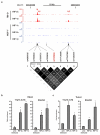
Figure 1
Figure 1. HIF-binding at 11q13.3
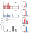
(a) Genome Browser tracks showing the read density at the 11q13.3 locus for HIF-2α and HIF-1β ChIP-seq in 786-O cells and for HIF-1α, HIF-2α and HIF-1β ChIP-seq in MCF-7 cells in which HIF-α chains were stabilized by hydroxylase inhibition. Three HIF-2-binding sites (H, h1 and h2) are identified in 786-O but not MCF-7 cells. Also shown are the positions of genotyped SNPs, together with LD analysis (r2 values). SNPs overlapping peak H and peak h2 are in sufficiently strong LD with the disease-associated tagSNP, rs7105934 (red) that the latter can act as a simple proxy. (b, c) ChIP-qPCR analysis confirming HIF-2α and HIF-1β binding at 11q13.3 (H) in both 786-0 cells (b) and renal tumor tissue (c); for comparison ChIP-qPCR at an established HIF-binding site at the EGLN3 locus, is shown. Bars show mean ± SD (n=3); *p<0.05, compared to pre-immune serum.
Figure 2
Figure 2. Chromatin structure and function of an enhancer at 11q13.3
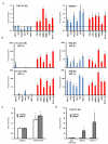
(a) Formaldehyde-assisted isolation of regulatory elements (FAIRE) was performed in 786-O and MCF-7 cells to map nucleosome occupancy at the 11q13.3 locus. qPCR products were normalized to those obtained for input DNA at this locus and then related to values obtained at a nearby control region outside the putative enhancer (ctrl, far right bar). Data is plotted against chromosomal location; the position of the ChIP-seq signals for HIF-1β and HIF-2α is illustrated below. (b-e) ChIP-qPCR signals at 11q13.3 (H) obtained from 786-0 cells (red) or MCF-7 cells (blue) using antibodies against (b) H3K4me1, (c) H3K4me3, (d) H3K27ac and (e) RNApol2; for comparison signals at a ubiquitous HIF-binding enhancer (NDRG1, Supplemental Fig. 3b), a HIF-binding promoter (FAM13A1) and a nearby non-enhancer control region are shown. (f) Luciferase reporter assay performed in 786-O cells using the sequences 11q13.3 (H) or 11q13.3 (H+h1) (indicated above); mut indicated mutation of the core HRE motif in H. Data is normalized to co-transfected β-galactosidase activity, mean ± SD (n=3).
Figure 3
Figure 3. Cell-type specificity of the 11q13.3 enhancer
(a) FAIRE analysis; data is shown for a panel of six pVHL wild-type cell lines (blue) in which HIF was induced using HIF hydroxylase inhibitor dimethyl oxalylglycine (DMOG) and six pVHL-defective cell lines (red). qPCR products were normalized as described. Data in the left panel is the enrichment value for primers centered on the ChIP-seq signal (H); for comparison signals at the ubiquitous HIF-binding enhancer (NDRG1) are also given (right panel). (b) ChIP-qPCR results from the same panel of pVHL wild-type and pVHL-defective cell lines for HIF-2α (upper) and HIF-1β binding (lower) at 11q13.3 (H) (left panel); for comparison signals at the EGLN3 locus are shown (right panels) (c) FAIRE analysis in 786-O cells re-expressing wild type pVHL (786-0/VHL cells). Data is the enrichment value for primers centered on the ChIP-seq signal (H) normalized to the control (non-enhancer) site; cells treated with or without DMOG to induce HIF (d) ChIP-qPCR analysis for HIF-2α and HIF-1β at the 11q13.3 H peak in 786-0/VHL cells treated with or without DMOG. Data is mean ± SD (n=3); *p<0.05 relative to pre-immune (ChIP) or to a control site (FAIRE).
Figure 4
Figure 4. Physical association of the remote HIF-binding site with the CCND1 promoter
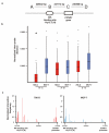
(a) Location and distance between midpoints of fosmid probes used for 3-dimensional (3D) fluorescent in situ hybridization (FISH). (b) Box-and-whisker plots showing median, inter-quartile range and full range of visualized inter-probe distances for each pair of probes, normalized to nuclear volume. Kruskal-Wallis 1-way ANOVA was significant (P<0.001). Mann-Whitney U-test showed that probes at the HIF-binding site and the CCND1 gene were significantly (*, p<0.001) more closely associated (in 786-O, but not MCF-7 cells) than were other probe pairs. (c) Chromatin conformation capture (3C) assays using the remote enhancer site as the anchor. Bars show qPCR analysis of the re-ligation frequency following restriction enzyme digestion, for cellular DNA, normalized to that of the BAC clones spanning the region of interest. Sites at the 11q13.3 HIF-binding locus preferentially ligated with fragments at the CCND1 promoter in 786-O cells, but not in MCF-7 cells indicated a cell-type specific physical interaction.
Figure 5
Figure 5. The minor (RCC-protective) allele at 11q13.3 disrupts HIF-binding and enhancer activity
(a) Genotype-specific PCR assays at the rs7948643 SNP, performed on immunoprecipitated chromatin from KTCL140 cells. The x-axis shows the intensity of the PCR signal for the minor (protective, a) and the y-axis shows intensity for the major (predisposing, A) allele. Control DNA from individuals homozygous (AA, aa) or heterozygous (Aa) for each allele was included for comparison in every assay. Input material and chromatin immunoprecipitated with pre-immune serum (control) showed an allelic ratio comparable to heterozygous DNA. Preferential recovery of allele A by HIF-2α and HIF-1β ChIP (yellow and black) demonstrates preferential binding of both subunits to this allele. (b) Genotype specific PCR assays at the rs7948643 SNP, performed on DNA immunoprecipitated from KTCL140 cells using an antibody directed against RNApol2 shows greater interaction of allele A with RNApol2. (c) Genotype specific PCR assays at the rs7948643 SNP, performed on KTCL140 DNA prepared by FAIRE. Preferential recovery of allele A indicates greater DNA accessibility. (d and e) Genotype specific PCR assay for rs7177, which lies in the 3′UTR of the CCND1 gene using (d) cDNA from KTCL140 cells or (e) immunoprecipitated DNA using an antibody against RNApol2 show allele specific expression of cyclin D1. Genomic DNA from KTCL140 or control DNA from individuals homozygous (BB, bb) or heterozygous (Bb) for each allele was included for comparison. Each symbol represents an individual PCR assay.

Comment in

References

    1. Purdue MP, et al. Genome-wide association study of renal cell carcinoma identifies two susceptibility loci on 2p21 and 11q13.3. Nat Genet. 2011;43:60–5. - PMC - PubMed
    1. Ferlay J, et al. Estimates of worldwide burden of cancer in 2008: GLOBOCAN 2008. Int J Cancer. 2010;127:2893–917. - PubMed
    1. Gnarra JR, et al. Mutations of the VHL tumour suppressor gene in renal carcinoma. Nat Genet. 1994;7:85–90. - PubMed
    1. Herman JG, et al. Silencing of the VHL tumor-suppressor gene by DNA methylation in renal carcinoma. Proc Natl Acad Sci U S A. 1994;91:9700–4. - PMC - PubMed
    1. Maxwell PH, et al. The tumour suppressor protein VHL targets hypoxia-inducible factors for oxygen-dependent proteolysis. Nature. 1999;399:271–5. - PubMed

References for Methods section

    1. Mole DR, et al. Genome-wide association of hypoxia-inducible factor (HIF)-1alpha and HIF-2alpha DNA binding with expression profiling of hypoxia-inducible transcripts. J Biol Chem. 2009;284:16767–75. - PMC - PubMed
    1. Brown J, et al. Subtelomeric chromosome rearrangements are detected using an innovative 12-color FISH assay (M-TEL) Nat Med. 2001;7:497–501. - PubMed
    1. Brown JM, et al. Coregulated human globin genes are frequently in spatial proximity when active. J Cell Biol. 2006;172:177–87. - PMC - PubMed
    1. Abramoff MD, Magalhaes PJ, Ram SJ. Image Processing with ImageJ. Biophotonics International. 2004;11:36–42.
    1. Hagege H, et al. Quantitative analysis of chromosome conformation capture assays (3C-qPCR) Nat Protoc. 2007;2:1722–33. - PubMed

Publication types

MeSH terms

Substances

Associated data