. 2013 Sep 26;155(1):107-20.
doi: 10.1016/j.cell.2013.08.061.

Hira-dependent histone H3.3 deposition facilitates PRC2 recruitment at developmental loci in ES cells

Affiliations

Hira-dependent histone H3.3 deposition facilitates PRC2 recruitment at developmental loci in ES cells

Laura A Banaszynski et al. Cell. .

Abstract

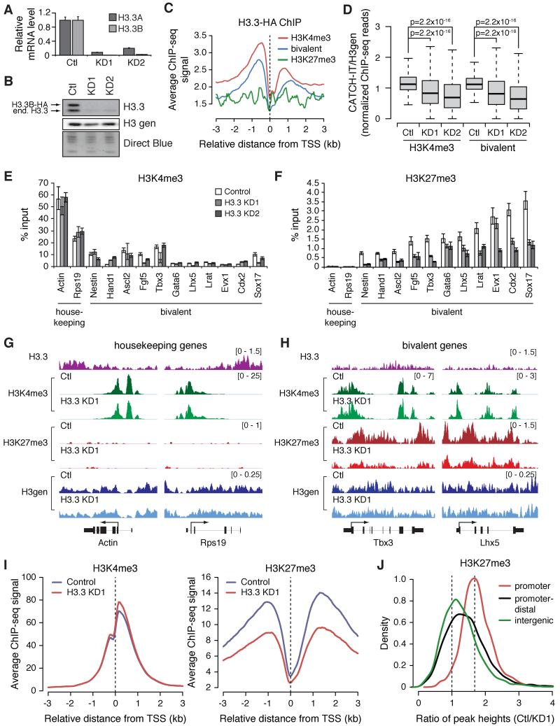
Polycomb repressive complex 2 (PRC2) regulates gene expression during lineage specification through trimethylation of lysine 27 on histone H3 (H3K27me3). In Drosophila, polycomb binding sites are dynamic chromatin regions enriched with the histone variant H3.3. Here, we show that, in mouse embryonic stem cells (ESCs), H3.3 is required for proper establishment of H3K27me3 at the promoters of developmentally regulated genes. Upon H3.3 depletion, these promoters show reduced nucleosome turnover measured by deposition of de novo synthesized histones and reduced PRC2 occupancy. Further, we show H3.3-dependent interaction of PRC2 with the histone chaperone, Hira, and that Hira localization to chromatin requires H3.3. Our data demonstrate the importance of H3.3 in maintaining a chromatin landscape in ESCs that is important for proper gene regulation during differentiation. Moreover, our findings support the emerging notion that H3.3 has multiple functions in distinct genomic locations that are not always correlated with an "active" chromatin state.


Figures

Figure 1
Figure 1. H3.3 Depletion Results in Global Loss of Nucleosome Turnover and Reduced H3K27me3 Enrichment at Promoters of Developmentally Regulated Genes
A) H3.3 transcript levels in control and H3.3-depleted ESCs (n = 3 ± s.d.). B) Immunoblot of H3.3 and total H3 in nuclear extracts from control and H3.3-depleted ESCs. Direct blue stain as loading control. C) H3.3 enrichment profiles at the transcription start site (TSS) of genes classified by bivalency (Mikkelsen et al., 2007). D) Box plots represent CATCH-IT normalized to H3gen ChIP-seq at promoters in control and H3.3 KD ESCs. The bottom and top of the boxes correspond to the 25th and 75th percentiles, and the internal band is the 50th percentile (median). The plot whiskers correspond to 1.5× interquartile range. P-values were calculated using Wilcoxon tests. ChIP-qPCR analyses of (E) H3K4me3 and (F) H3K27me3 at representative housekeeping and bivalent genes in control and H3.3-depleted ESCs. Error bars represent s.d. from one experiment (n = 3). Data are representative of at least 3 independent ChIP experiments. Genome browser representations of H3.3, H3K4me3, H3K27me3, and H3gen profiles in control and H3.3 KD1 ESCs for (G) two housekeeping genes, Actin (left) and Rps19 (right), and (H) two developmentally regulated bivalent genes in ESCs, Tbx3 (left) and Lhx5 (right). Read counts normalized to total number of reads for each data set. I) ChIP-seq profiles of H3K4me3 (left) and H3K27me3 (right) at the TSS of bivalent genes in control and H3.3 KD1 ESCs. Data are represented as reads per mapped million per 50 bp bin. J) Ratio of H3K27me3 peak heights in control vs. H3.3 KD1 ESCs at promoter (±2 kb from TSS), promoter-distal (−50 kb from TSS to +5 kb from transcription end site (TES) excluding promoter peaks), and intergenic (all other) peaks, defined by distance from nearest annotated genes. X-axis values >1 indicate reduced H3K27me3 enrichment upon H3.3 depletion. See also Figure S1 and Table S1.
Figure 2
Figure 2. Transcription Factors Essential for Trophectoderm Specification are Upregulated upon H3.3 Depletion
RNA-seq data sets were obtained from control and H3.3 KD ESC poly(A)-RNA, and fragments per kilobase per million mapped reads (FPKM) were calculated for all mouse Ensembl genes. Differentially expressed genes were identified using Cuffdiff with FDR < 0.05. FPKMs for genes (A) upregulated or (B) downregulated in both H3.3 KD1 and H3.3 KD2 (Venn diagrams) with respect to control are represented by box plots. The bottom and top of the boxes correspond to the 25th and 75th percentiles, and the internal band is the 50th percentile (median). The plot whiskers correspond to the 10th and 90th percentiles. P-values were calculated using Wilcoxon tests. C) Gene ontology biological process categories for genes significantly upregulated upon H3.3 knockdown from analysis performed with DAVID software (Huang et al., 2009). D) RT-qPCR of pluripotency and selected lineage marker genes in control and H3.3 KD ESCs. Data are represented as mean expression relative to Gapdh with control normalized to 1. Error bars represent standard deviation (s.d.) for an experiment performed in biological triplicate and technical duplicate, ***p<0.001. E) Immunoblot of control ESCs and H3.3 KD1 ESCs exogenously expressing C-terminally Flag-HA tagged H3.2 or H3.3. F) RT-qPCR of trophectoderm-specific transcription factors in control ESCs and H3.3 KD1 ESCs exogenously expressing either H3.2 or H3.3. Data are represented as mean expression relative to Gapdh ± s.d. (n = 3). Box plots representing ChIP-seq read counts at promoters of genes upregulated by RNA-seq (RNA-seq up) and bivalent promoters for (G) H3K4me3, (H) H3K27me3, and (I) H3K27ac in both control and H3.3 KD1 ESCs. Plots as in panels A and B, except plot whiskers correspond to 1.5× interquartile range. See also Figure S2 and Table S2.
Figure 3
Figure 3. H3.3-Depleted ESCs Show Altered Potential and Misregulation of Lineage-Specific Factors Upon Differentiation
A) Histology of teratomas from control (left panel) and H3.3 KD1 (right panel) ESCs. Sections were stained with Hematoxylin and Eosin (H&E). Trophoblast focus outlined and representative giant trophoblast cells indicated by arrows (right panel). B) RT-qPCR of placental markers in control and H3.3 KD1 teratomas. Data are represented as mean expression relative to Gapdh ± s.d. for an experiment performed in biological and technical triplicate. *p<0.05, **p<0.01, ***p<0.001. C-E) RT-qPCR of indicated lineage markers in control and H3.3 KD1 ESCs subject to differentiation. Data are represented as mean expression relative to Gapdh ± s.d. (n = 3) at time points indicated, normalized to control ESCs. F) Fold-enrichment over ESC of genes defined as ESC-bivalent found differentially regulated in H3.3-null MEFs. For all comparisons, p<1x10⁻⁶. See also Figure S3.
Figure 4
Figure 4. H3.3 Contributes to PRC2 Binding at Bivalent Promoters
A) ChIP-qPCR of Suz12 (top) and Jarid2 (bottom) at representative housekeeping and bivalent genes in control and H3.3-depleted ESCs. Error bars represent s.d. (n = 3). Data confirmed by multiple biological replicates. B) H3.3 enrichment profile at the TSS of bivalent genes in wild type and Suz12−/− ESCs. Data are represented as reads per mapped million per 50 bp bin. C) RT-qPCR of trophectoderm-specific transcription factors in control ESCs and H3.3 KD1 ESCs exogenously expressing H3.2, H3.3, or H3.3 mutated at K27. Data are represented as mean expression relative to Gapdh ± s.d. (n = 3). Data from control and H3.2/H3.3 addback are also shown in Figure 2F. See also Figure S4.
Figure 5
Figure 5. Maintenance of H3K27me3 Levels at Bivalent Promoters Requires Hira-Dependent H3.3 Deposition
A-D) ChIP-qPCR of (A, B) H3K4me3 and (C, D) H3K27me3 at representative housekeeping and bivalent genes in (A, C) control and Hira−/− ESCs and (B, D) control and Daxx-depleted ESCs. ChIP-qPCR of (E) Suz12 and (F) Jarid2 at representative housekeeping and bivalent genes in control and Hira−/− ESCs. For panels A-F, error bars represent s.d. (n=3) with data confirmed by 3 biological replicates. G) Box plots represent CATCH-IT normalized to H3gen ChIP-seq at promoters in control and Hira−/− ESCs. The bottom and top of the boxes correspond to the 25th and 75th percentiles, and the internal band is the 50th percentile (median). The plot whiskers correspond to 1.5× interquartile range. P-values were calculated using Wilcoxon tests. H) RT-qPCR of trophectoderm markers in control and Hira−/− ESCs subject to differentiation. Data are represented as mean expression relative to Gapdh ± s.d. (n = 3) at the time points indicated, normalized to control ESCs. See also Figure S5.
Figure 6
Figure 6. Hira Co-localizes with Promoter-proximal RNAPII and PRC2 at Bivalent Promoters in an H3.3-dependent Manner
A) Genome browser representation of input, H3.3, and Hira profiles for chromosome 11. Read counts are normalized to total number of reads obtained for each data set. B) ChIP-seq density heat maps for RefSeq genes classified as H3K4me3 only (n = 15,670), bivalent (n = 2,938), or H3K27me3 only (n = 23). Plots show ±5 kb centered on the TSS. H3K4me3 only genes are rank-ordered by H3K4me3 enrichment, and bivalent and H3K27me3 only genes are rank-ordered by H3K27me3 enrichment, based on FPKM for each ChIP-seq data set. Data are represented as reads per mapped million per 50 bp bin. Color intensity represents tag counts, standardized across gene groupings for each ChIP-seq data set. C) Immunoblotting of Hira immunoprecipitated from wild type or H3.3-null nuclear extracts showing coimmunoprecipitation with Ubinuclein and H3.3 (* non-specific band) (5% input), and H3.3-dependent coimmunoprecipitation with RNAPII S5p, Ezh2, Suz12, and Jarid2 (0.5% input). D) Hira enrichment profile at the TSS of genes classified by chromatin modification state in wild type (solid lines) and H3.3 KO2 (dashed lines) ESCs. Data are represented as reads per mapped million per 50 bp bin. E) Model of H3.3 in modulation of PRC2 recruitment and activity in ESC. Recruitment of Hira and H3.3 to PRC2 targets are co-dependent and upstream of PRC2 recruitment. Either Hira or H3.3 directly, or the high frequency of free DNA afforded by their presence, allow for optimal PRC2 recruitment and activity at target loci. See also Figure S6.

References

    1. Bernstein BE, Mikkelsen TS, Xie X, Kamal M, Huebert DJ, Cuff J, Fry B, Meissner A, Wernig M, Plath K, et al. A bivalent chromatin structure marks key developmental genes in embryonic stem cells. Cell. 2006;125:315–326. - PubMed
    1. Brookes E, de Santiago I, Hebenstreit D, Morris KJ, Carroll T, Xie SQ, Stock JK, Heidemann M, Eick D, Nozaki N, et al. Polycomb Associates Genome-wide with a Specific RNA Polymerase II Variant, and Regulates Metabolic Genes in ESCs. Cell Stem Cell. 2012;10:157–170. - PMC - PubMed
    1. Brookes E, Pombo A. Modifications of RNA polymerase II are pivotal in regulating gene expression states. EMBO Rep. 2009;10:1213–1219. - PMC - PubMed
    1. Bush KM, Yuen BT, Barrilleaux BL, Riggs JW, O’Geen H, Cotterman RF, Knoepfler PS. Endogenous mammalian histone H3.3 exhibits chromatin-related functions during development. Epigenetics Chromatin. 2013;6:7. - PMC - PubMed
    1. Couldrey C, Carlton MB, Nolan PM, Colledge WH, Evans MJ. A retroviral gene trap insertion into the histone 3.3A gene causes partial neonatal lethality, stunted growth, neuromuscular deficits and male sub-fertility in transgenic mice. Hum Mol Genet. 1999;8:2489–2495. - PubMed

Publication types

MeSH terms

Associated data