. 2014 Apr 7:5:3615.
doi: 10.1038/ncomms4615.

Lactate racemase is a nickel-dependent enzyme activated by a widespread maturation system

Affiliations

Lactate racemase is a nickel-dependent enzyme activated by a widespread maturation system

Benoît Desguin et al. Nat Commun. .

Abstract

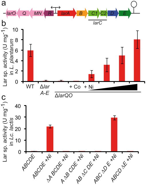
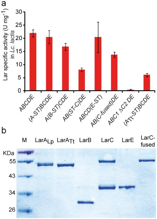
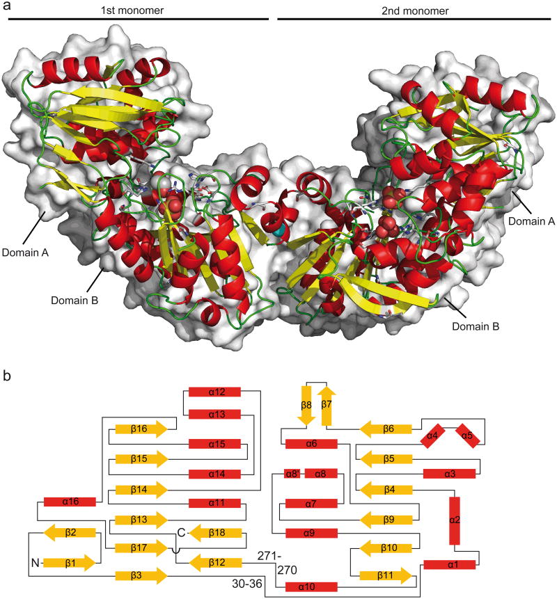
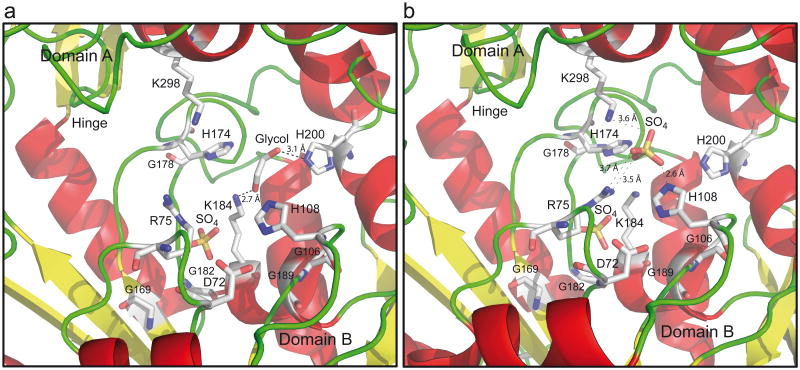
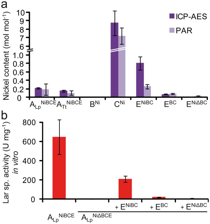
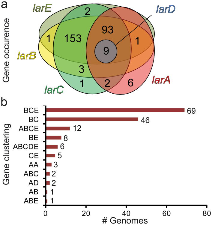
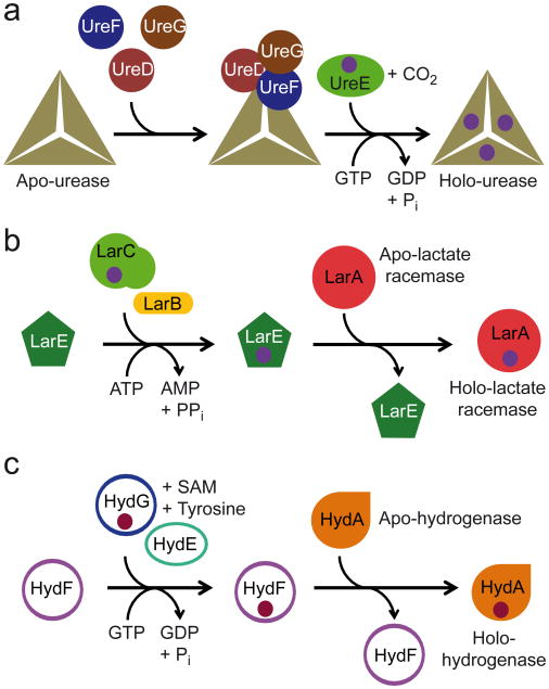
Racemases catalyse the inversion of stereochemistry in biological molecules, giving the organism the ability to use both isomers. Among them, lactate racemase remains unexplored due to its intrinsic instability and lack of molecular characterization. Here we determine the genetic basis of lactate racemization in Lactobacillus plantarum. We show that, unexpectedly, the racemase is a nickel-dependent enzyme with a novel α/β fold. In addition, we decipher the process leading to an active enzyme, which involves the activation of the apo-enzyme by a single nickel-containing maturation protein that requires preactivation by two other accessory proteins. Genomic investigations reveal the wide distribution of the lactate racemase system among prokaryotes, showing the high significance of both lactate enantiomers in carbon metabolism. The even broader distribution of the nickel-based maturation system suggests a function beyond activation of the lactate racemase and possibly linked with other undiscovered nickel-dependent enzymes.


Figures

Figure 1
Figure 1. Analysis of the lar gene cluster and its encoded Lar proteins
(a) lar locus of L. plantarum with the two operons larR(MN)QO and larA-E. (b) Effect of larA-E and lar(MN)QO deletions in L. plantarum on Lar specific activity in crude extracts. Supplementation assays of the ΔlarQO mutant with CoCl2 (1 mM) and NiCl2 (0.2, 0.4, 0.8 and 1.5 mM). (c) Lar specific activity in crude extracts after the expression of lar genes in Lc. lactis: larA-E operon +/- NiCl2 (1 mM), in frame deletions of individual genes (Δ) in the larA-E operon with NiCl2 supplementation (1 mM). Data in panels b and c are average of quadruplicates from one representative experiment of three independent experiments showing similar results. The error bars represent the 95% confidence interval (Student's t test).
Figure 2
Figure 2. StrepII-tag insertions in the larA-E operon and LarC modifications
(a) Lactate racemase specific activity of Lc. lactis strains expressing the entire larA-E operon of L. plantarum (ABCDE) in which one of the Lar proteins has been fused to a StrepII-tag at either the N- (ST-X) or C-terminus (X-ST), harbouring a 1 bp insertion at the end of larC1 (C-fused), with the in frame deletions of larC2 (ΔC2) and expressing the artificial operon larATtBCDE in which LarATt has been fused to a StrepII-tag at the C-terminus (ATt-ST). NiCl2 was added in all cases (1mM). Data are average of quadruplicates from one representative experiment of two independent experiments showing similar results. The error bars represent the 95% confidence interval (Student's t test). (b) SDS-PAGE of purified LarALp, LarATt, LarB, LarC (LarC1 and LarC1C2), LarE, and LarC-fused (only LarC1C2).
Figure 3
Figure 3. 3D structure and topology of LarA
(a) 3D dimeric structure of LarATt (RSCB Protein Data Bank (PDB) accession code 2YJG). α-Helices are in red, β-sheets in yellow, and loops in green. Conserved residues are shown in stick representation and ethylene glycol, sulfate and Mg(II) are in sphere representation with C in white, N in blue, O in red, Mg in cyan and S in yellow. Surface representation is shown at the background. (b) The lactate racemase fold. α-Helices are in red, β-strands in yellow and loops are in black. The topology of the β-sheet of domain A is six parallel β-strands in the order 162345. The topology of the β-sheet of domain B is six β-strands in the order 321456 with the last β-strand antiparellel to the rest. The numbers indicate the position of the two hinges.
Figure 4
Figure 4. LarA catalytic site
(a) Catalytic site of the first monomer (b) Catalytic site of the second monomer. Sulfates and ethylene glycol are displayed. Color code as in Fig. 3a.
Figure 5
Figure 5. XANES and EXAFS spectra
(a) Ni K-edge XANES spectra of LarATt and LarALp showing a four square planar or five square pyramidal coordination geometry for nickel binding; 10 mM Tris buffer, pH 7.5, 20% glycerol. (b) Ni K-edge EXAFS spectra of LarATt and fit for N2H1H1S1. Fourier transformed EXAFS spectra (no phase correction, FT window = 2-12.5 Å-1). Inset: k3-weighted unfiltered EXAFS spectra; data (circles), best fit (line). (c) Ni K-edge EXAFS spectra of LarATt and fit for GH2H1S1. Fourier transformed EXAFS spectra (no phase correction, FT window = 2-12.5 Å-1). Inset: k3-weighted unfiltered EXAFS spectra; data (circles), best fit (line). N: N/O scatterers, H: histidine scatterer, S: S/Cl scatterers, and G: glycerol scatterer.
Figure 6
Figure 6. Lar accessory proteins
(a) Ni content of Lar proteins as measured by PAR assays and ICP-AES. PAR data are average of two independent experiments (triplicates in each experiment). ICP-AES data are average of duplicates from one experiment. (b) Specific Lar activity of purified proteins and in vitro activation of apo-LarA. ALpNiBCE, LarA purified from a Lc. lactis strain expressing LarBCE and cultivated in the presence of Ni(II); ALpNiΔBCE, LarA purified from a Lc. lactis strain not expressing LarBCE and cultivated in the presence of Ni(II); +ENiBC, assay performed in the presence of an excess of LarE purified from a Lc. lactis strain expressing LarBC and cultivated in the presence of Ni(II); +EBC, assay performed in the presence of an excess of LarE purified from a Lc. lactis strain expressing LarBC and cultivated in the absence of Ni(II); +ENiΔBC, assay performed in the presence of an excess of LarE purified from a Lc. lactis strain not expressing LarBC and cultivated in the presence of Ni(II); BNi, LarB purified from a Lc. lactis strain cultivated in the presence of Ni(II); and CNi, LarC purified from a Lc. lactis strain cultivated in the presence of Ni(II). Data are average of quadruplicates from one representative experiment of two independent experiments showing similar results. L-lactate was used as a substrate for all Lar activity measurements. The error bars represent the 95% confidence interval (Student's t test) in panels a and b.
Figure 7
Figure 7. In silico analysis of lar genes
(a) Distribution of the lar genes in 1087 sequenced bacterial and archaeal genomes. The Venn diagram illustrates the occurrence and overlap of predicted larA (red), larB (yellow), larC (green), larD (blue) and larE genes (olive green). (b) Gene clustering of predicted larA (A), larB (B), larC (C), larD (D) and larE (E) genes in 1087 bacterial and archaeal genomes. The numbers indicate the total number of clusters of each type. Non-clustered representatives are not included in panel b.
Figure 8
Figure 8. Model of assembly of lactate racemase metallocenter
(a) Assembly of urease metallocenter, one triangle represents one urease trimer of UreABC. UreD, UreE, UreF, and UreG are the urease accessory proteins. (b) Proposed model for the assembly of lactate racemase metallocenter. LarA is the lactate racemase. LarB, LarC, and LarE are the lactate racemase accessory proteins. (c) Assembly of [FeFe]-hydrogenase metallocenter. HydA is the [FeFe]-hydrogenase. HydE, HydF, and HydG are the [FeFe]-hydrogenase accessory proteins. For clarity, the Fe-S clusters of [FeFe]-hydrogenase have been omitted. The purple balls represent Ni or a Ni-containing center in a and b. The dark red balls represent the H-cluster in c. ATP: adenosine triphosphate, AMP: adenosine monophosphate, GTP: guanosine triphosphate, GDP: guanosine diphosphate, PPi: pyrophosphate, Pi: phosphate, and SAM: S-adenosyl methionine.

References

    1. Okano K, Tanaka T, Ogino C, Fukuda H, Kondo A. Biotechnological production of enantiomeric pure lactic acid from renewable resources: recent achievements, perspectives, and limits. Appl Microbiol Biotechnol. 2010;85:413–423. - PubMed
    1. Thauer RK, Jungermann K, Decker K. Energy conservation in chemotrophic anaerobic bacteria. Bacteriol Rev. 1977;41:100–180. - PMC - PubMed
    1. Walsh CT. Vancomycin resistance: decoding the molecular logic. Science. 1993;261:308–309. - PubMed
    1. Goffin P, et al. Lactate racemization as a rescue pathway for supplying D-lactate to the cell wall biosynthesis machinery in Lactobacillus plantarum. J Bacteriol. 2005;187:6750–6761. - PMC - PubMed
    1. Garvie EI. Bacterial lactate dehydrogenases. Microbiol Rev. 1980;44:106–139. - PMC - PubMed

Publication types

MeSH terms

Associated data