. 2013 May;9(5):e1003489.
doi: 10.1371/journal.pgen.1003489. Epub 2013 May 2.

Female bias in Rhox6 and 9 regulation by the histone demethylase KDM6A

Affiliations

Female bias in Rhox6 and 9 regulation by the histone demethylase KDM6A

Joel B Berletch et al. PLoS Genet. 2013 May.

Abstract

The Rhox cluster on the mouse X chromosome contains reproduction-related homeobox genes expressed in a sexually dimorphic manner. We report that two members of the Rhox cluster, Rhox6 and 9, are regulated by de-methylation of histone H3 at lysine 27 by KDM6A, a histone demethylase with female-biased expression. Consistent with other homeobox genes, Rhox6 and 9 are in bivalent domains prior to embryonic stem cell differentiation and thus poised for activation. In female mouse ES cells, KDM6A is specifically recruited to Rhox6 and 9 for gene activation, a process inhibited by Kdm6a knockdown in a dose-dependent manner. In contrast, KDM6A occupancy at Rhox6 and 9 is low in male ES cells and knockdown has no effect on expression. In mouse ovary where Rhox6 and 9 remain highly expressed, KDM6A occupancy strongly correlates with expression. Our study implicates Kdm6a, a gene that escapes X inactivation, in the regulation of genes important in reproduction, suggesting that KDM6A may play a role in the etiology of developmental and reproduction-related effects of X chromosome anomalies.


Conflict of interest statement

The authors have declared that no competing interests exist.

Figures

Figure 1
Figure 1. Sexual dimorphism of Rhox6 and 9 and Kdm6a expression in ES cells and embryos.
(A) Rhox6 and 9 expression measured by qRT-PCR is higher in female (PGK12.1 and E8) than male (WD44 and E14) undifferentiated ES cells (***p<0.0001). Gene expression was normalized to 18s levels. (B) Rhox6 and 9 expression measured by qRT-PCR in female PGK12.1 ES cells and in male WD44 ES cells during ES cell differentiation. Gene expression was normalized to 18s levels. (C) Re-analyses of published expression array data in germ cells and somatic cells in sexed embryos (11.5–13.5 dpc) shows higher Rhox6 and 9 expression in female than male embryos (*p<0.05, **p<0.001) (see also Figure S2A). Endothelial, mesenchymal, and follicle cells were analyzed together as somatic cells (12 samples total). Values were normalized to the array mean. (D) Kdm6a expression measured by qRT-PCR is higher in female (PGK12.1 and E8) than male (WD44 and E14) undifferentiated ES cells (*p<0.05). Gene expression was normalized to 18s levels. (E) Western blot analysis confirms higher protein levels in female (PGK12.1 and E8) versus male (WD44 and E14) ES cells. β-ACTIN is used as a control. (F) Kdm6a expression measured by qRT-PCR in female PGK12.1 ES cells and in male WD44 ES cells is higher in undifferentiated female than male ES cells throughout differentiation (**p<0.001, ***p<0.0001). Gene expression was normalized to 18s levels. (G) Re-analyses of published expression array data in germ cells and somatic cells in sexed embryos (11.5–13.5 dpc) shows higher Kdm6a expression in female than male embryos (*p<0.05, **p<0.001, ***p<0.0001). Endothelial, mesenchymal, and follicle cells were analyzed together as somatic cells (12 samples total). was higher. Values were normalized to the array mean.
Figure 2
Figure 2. KDM6A is preferentially recruited to Rhox6 and 9 in female ES cells.
(A) ChIP-qPCR analysis of KDM6A occupancy at the 5′ end of Rhox6 and 9 is higher in female (PGK12.1 and E8) than male (WD44 and E14) undifferentiated ES cells (*p<0.05). (B) H3K4me3 enrichment during differentiation of female PGK12.1 and male WD44 ES cells shows lower levels in male ES cells and a decrease of between day 0 and 15 in agreement with gene silencing after differentiation of these ES cells (see also Figure 1). (C) KDM6A occupancy at the 5′ end of Rhox6 and 9 during differentiation of female PGK12.1 ES cells and male WD44 ES cells. (D) H3K27me3 levels at the 5′ end of Rhox6 and 9 mirror KDM6A occupancy changes. The increase at day 15 is due to X inactivation in female PGK12.1 ES cells (see also Figures S2B, S4, and S6). Average enrichment/occupancy for two separate ChIP experiments is shown as ChIP/input (A, B, C).
Figure 3
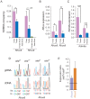
Figure 3. Kdm6a knockdown causes a female-specific decrease in Rhox6 and 9 expression in ES cells.
(A) Quantitative RT-PCR after Kdm6a knockdown in female PGK12.1 ES cells shows a 75% and a 52% decrease in Kdm6a and Rhox6 and 9 expression, respectively. Expression is shown relative to control levels obtained with scrambled siRNA. Control gene β-actin levels are set to 1. The inset shows a western blot using two different KDM6A antibodies, which confirms a ∼70–90% reduction in protein levels. β-ACTIN is used as a control. (B) Kdm6a knockdown results in a decrease in Rhox6 and 9 expression in female (PGK12.1 and E8) but not male (WD44 and E14) undifferentiated ES cells. Expression measured by array analysis (PGK12.1 and WD44) and qRT-PCR (E8 and E14) is shown as fold change compared to the control gene β-actin. Array results are from four independent experiments and qRT-PCR results are from three independent experiments. (C) The decrease in Rhox6 and 9 expression correlates with the level of Kdm6a knockdown in a dose dependent manner in PGK12.1 ES cells (*p<0.05). Fold changes in levels of Rhox6 and 9 measured by expression arrays are shown relative to fold changes in Kdm6a levels. (D) Kdm6a knockdown results in a significant (*p<0.05) increase in H3K27me3 enrichment at Rhox6 and 9 5′end, gene body, and 3′end. ChIP-qPCR of H3K27me3 enrichment in PGK12.1 ES cells treated with control scrambled siRNA and Kdm6a specific siRNA. Average enrichment for two separate ChIP experiments is shown as ChIP/input.
Figure 4
Figure 4. Rhox6 and 9 are bivalent and preferentially occupied by KDM6A in female ES cells.
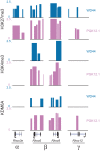
H3K27me3, H3K4me3 and KDM6A enrichment profiles in undifferentiated female PGK12.1 (pink) and male WD44 ES (blue) cells at representative genes from each Rhox subcluster (α, β, and γ) demonstrate that only Rhox6 and 9 are highly enriched with both histone modifications and are bound by KDM6A (see also Figure S6). Rhox3e (α cluster) is enriched in H3K27me3 but not H3K4me3 or KDM6A, and Rhox12 (γ cluster) shows little enrichment for the proteins analyzed. Significant enrichment peaks based on Nimblescan analysis (FDR score <.05) are shown. Data uploaded to UCSC genome browser (NCBI36/mm8).
Figure 5
Figure 5. Rhox6 and 9 expression and KDM6A occupancy are high in ovary where the genes are imprinted.
(A) Rhox6 and 9 have significantly higher expression in mouse ovary than in testis, based on re-analyses of published expression array data for 14 testis and 12 ovary specimens (*p<0.05, **p<0.001). Expression normalized to array mean (see also Figure 1C). (B) KDM6A occupancy measured by ChIP-qPCR at the 5′end of Rhox6 and 9 is higher in ovary than in testis, and is very low to undetectable in brain where these genes are not expressed . Occupancy levels were normalized to input fractions. (C) Kdm6a has high expression in female tissues especially ovary based on analyses of published expression array data (***p<0.0001) (see also Figure 1F). (D) Rhox6 and 9 are expressed from the maternal allele only in ovary because of imprinting. DNA sequence chromatograms of gDNA and RT-PCR (cDNA) products derived from ovary from female F1 mice obtained by mating M. spretus males with C57BL/6J females with or without an Xist mutation (XistΔ and XistΔ−). SNPs to distinguish Rhox6 and 9 alleles on the active X (Xa) and on inactive X (Xi) are indicated below. In ovary from both XistΔ and XistΔ− mice the gDNA shows heterozygosity at the SNPs while the cDNA shows only the maternal allele, consistent with paternal imprinting. (E) By qRT-PCR Rhox6 and 9 are more highly expressed in ovary from XistΔ mice in which the maternal X chromosome is expressed in all cells, compared to XistΔ− mice in which there is random X inactivation (1.7-fold and 3-fold, respectively), suggesting that Rhox6 and 9 are silenced by X inactivation. Values represent the expression ratio between XistΔ and XistΔ− ovaries.

References

    1. Weatherbee SD, Halder G, Kim J, Hudson A, Carroll S (1998) Ultrabithorax regulates genes at several levels of the wing-patterning hierarchy to shape the development of the Drosophila haltere. Genes Dev 12: 1474–1482. - PMC - PubMed
    1. Wang J, Mager J, Schnedier E, Magnuson T (2002) The mouse PcG gene eed is required for Hox gene repression and extraembryonic development. Mamm Genome 13: 493–503. - PubMed
    1. Lan F, Bayliss PE, Rinn JL, Whetstine JR, Wang JK, et al. (2007) A histone H3 lysine 27 demethylase regulates animal posterior development. Nature 449: 689–694. - PubMed
    1. Swigut T, Wysocka J (2007) H3K27 demethylases, at long last. Cell 131: 29–32. - PubMed
    1. Cho YW, Hong T, Hong S, Guo H, Yu H, et al. (2007) PTIP associates with MLL3- and MLL4-containing histone H3 lysine 4 methyltransferase complex. J Biol Chem 282: 20395–20406. - PMC - PubMed

Publication types

MeSH terms

Associated data