. 2013 May 31;8(5):e64800.
doi: 10.1371/journal.pone.0064800. Print 2013.

Bcl11a controls Flt3 expression in early hematopoietic progenitors and is required for pDC development in vivo

Affiliations

Bcl11a controls Flt3 expression in early hematopoietic progenitors and is required for pDC development in vivo

Xiaodi Wu et al. PLoS One. .

Abstract

Bcl11a is a transcription factor known to regulate lymphoid and erythroid development. Recent bioinformatic analysis of global gene expression patterns has suggested a role for Bcl11a in the development of dendritic cell (DC) lineages. We tested this hypothesis by analyzing the development of DC and other lineages in Bcl11a (-/-) mice. We found that Bcl11a was required for expression of IL-7 receptor (IL-7R) and Flt3 in early hematopoietic progenitor cells. In addition, we found severely decreased numbers of plasmacytoid dendritic cells (pDCs) in Bcl11a (-/-) fetal livers and in the bone marrow of Bcl11a (-/-) fetal liver chimeras. Moreover, Bcl11a (-/-) cells showed severely impaired in vitro development of Flt3L-derived pDCs and classical DCs (cDCs). In contrast, we found normal in vitro development of DCs from Bcl11a (-/-) fetal liver cells treated with GM-CSF. These results suggest that the persistent cDC development observed in Bcl11a (-/-) fetal liver chimeras reflects derivation from a Bcl11a- and Flt3-independent pathway in vivo.


Conflict of interest statement

Competing Interests: The authors have declared that no competing interests exist.

Figures

Figure 1
Figure 1. Bcl11a is required for development of lymphoid and DC progenitors in the fetus.
(A) Flow cytometry analysis of progenitor populations in WT and Bcl11a −/− fetal livers dissected at embryonic day 14.5 (E14.5). Populations are gated as indicated; numbers represent the percentage of cells within the histogram that lie in the indicated gate. Data are representative of two mice per group. (B) Progenitor populations in WT and Bcl11a −/− fetal livers at E14.5, analyzed by flow cytometry as in (A) and presented as a percentage of total fetal liver cells. Bars represent the mean (± SEM) of two mice per group. (C) CD150 (Slamf1) expression within the LSK fraction in WT and Bcl11a −/− fetal livers at E14.5. (D) SL-GMPs in WT and Bcl11a −/− fetal livers at E14.5.
Figure 2
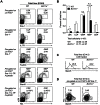
Figure 2. Bcl11a is required for development of lymphoid and DC progenitors in the adult.
(A) Flow cytometry analysis of progenitor populations in lethally irradiated congenic mice reconstituted with WT or Bcl11a −/− fetal liver cells, analyzed four weeks after transplant. Data are representative of three mice per group. (B) Progenitor populations in WT and Bcl11a −/− fetal liver chimeras at four weeks after transplant, analyzed by flow cytometry as in (A) and presented as a percentage of total BM cells. Bars represent the mean (± SEM) of three mice per group. (C) CD150 (Slamf1) expression within the donor-derived LSK fraction in WT and Bcl11a −/− fetal liver chimeras at four weeks after transplant. (D) SL-GMPs in WT and Bcl11a −/− fetal liver chimeras at four weeks after transplant.
Figure 3
Figure 3. Bcl11a regulates the expression of Flt3 and Il7r.
(A) Microarray analysis of sorted GMPs (left) and MPPs (right) from WT and Bcl11a −/− fetal liver chimeras. (B) Shown is a Venn diagram of probe sets (excluding normalization controls) with a greater than twofold change in expression between WT and Bcl11a −/− MPPs. (C) Shown are log2-transformed ratios of gene expression in Bcl11a −/− MPPs relative to WT MPPs (x-axis) plotted against log2-transformed ratios of gene expression in WT CDPs relative to WT monocytes (ImmGen; y-axis). For clarity, probe sets with less than twofold changes in expression (log2-transformed ratios between −1 and 1) along either dimension are omitted (gray). (D) Shown is a heat map of log2-transformed gene expression in WT and Bcl11a −/− GMPs and MPPs for probe sets that constitute an ImmGen core cDC signature. Highlighted are genes that show a greater than twofold change in expression between WT and Bcl11a −/− GMPs (red) or between WT and Bcl11a −/− MPPs (green).
Figure 4
Figure 4. Bcl11a is required in vivo for development of pDCs but not cDCs.
(A) Donor-derived (CD45.2+) chimerism in the BM, spleen, and skin-draining lymph node (SLN) of WT and Bcl11a −/− fetal liver chimeras. Bars represent the mean (± SEM) of three mice per group. (B) Flow cytometry analysis of donor-derived pDCs in the spleen. Data are representative of three mice per group. (C) Flow cytometry analysis of donor-derived cDCs in the spleen. Data are representative of three mice per group.
Figure 5
Figure 5. Bcl11a deficiency in vivo impairs development of lymphoid and myeloid populations.
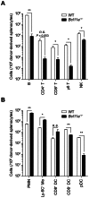
(A) Donor-derived lymphoid populations in the spleen of WT and Bcl11a −/− fetal liver chimeras, analyzed by flow cytometry. Bars represent the mean (± SEM) of three mice per group. (B) Donor-derived myeloid populations in the spleen of WT and Bcl11a −/− fetal liver chimeras, analyzed by flow cytometry as in Fig. 4. Bars represent the mean (± SEM) of three mice per group.
Figure 6
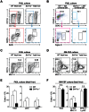
Figure 6. Bcl11a is required in vitro for development of Flt3L-derived pDCs and cDCs but not GM-CSF–derived cDCs.
(A) Flow cytometry analysis of pDCs in Flt3L cultures of fetal liver cells. Data are representative of three to four replicates over two experiments. (B) Flow cytometry analysis of pDCs in Flt3L cultures of BM cells derived from fetal liver chimeras. Data are representative of three replicates. (C, D) Flow cytometry analysis of Flt3L-derived cDCs (C) or GM-CSF-derived DCs (D) in cultures of fetal liver cells. Data are representative of three to four replicates over two experiments. (E, F) Counts of total cells and indicated subsets in Flt3L cultures (E) or GM-CSF cultures (F) of fetal liver cells, analyzed by flow cytometry as in (C) or (D), respectively. Bars represent the mean (± SEM) of three to four replicates per group pooled from two experiments.
Figure 7
Figure 7. Cytokine signaling in DC development and regulation by Bcl11a.
(A) Flow cytometry analysis of pDCs in WT and Il7r −/− spleens. Data are representative of four mice per group over two experiments. (B) Flow cytometry analysis of pDCs in WT and Flt3l −/− spleens. Data are representative of three mice per group over two experiments. (C) Flow cytometry analysis of donor-derived cDCs in the spleen of WT and Bcl11a −/− fetal liver chimeras, analyzed by flow cytometry as in Fig. 4. Data are representative of three mice per group. (D) Bcl11a binding in the Flt3 genomic locus assayed by ChIP-qPCR. Data are represented as fold enrichment as compared to isotype control.

References

    1. Naik SH, Metcalf D, van Nieuwenhuijze A, Wicks I, Wu L, et al. (2006) Intrasplenic steady-state dendritic cell precursors that are distinct from monocytes. Nat Immunol 7: 663–671. - PubMed
    1. Onai N, Obata-Onai A, Schmid MA, Ohteki T, Jarrossay D, et al. (2007) Identification of clonogenic common Flt3+ M-CSFR+ plasmacytoid and conventional dendritic cell progenitors in mouse bone marrow. Nature 8: 1207–1216. - PubMed
    1. Naik SH, Sathe P, Park HY, Metcalf D, Proietto AI, et al. (2007) Development of plasmacytoid and conventional dendritic cell subtypes from single precursor cells derived in vitro and in vivo. Nat Immunol 8: 1217–1226. - PubMed
    1. Liu K, Victora GD, Schwickert TA, Guermonprez P, Meredith MM, et al. (2009) In vivo analysis of dendritic cell development and homeostasis. Science 324: 392–397. - PMC - PubMed
    1. Wu L, Nichogiannopoulou A, Shortman K, Georgopoulos K (1997) Cell-autonomous defects in dendritic cell populations of Ikaros mutant mice point to a developmental relationship with the lymphoid lineage. Immunity 7: 483–492. - PubMed

Publication types

MeSH terms

Associated data