Comparative Study
. 2013 Dec;123(12):5135-45.
doi: 10.1172/JCI70231. Epub 2013 Nov 25.

Protein microarray analysis reveals BAFF-binding autoantibodies in systemic lupus erythematosus

Comparative Study

Protein microarray analysis reveals BAFF-binding autoantibodies in systemic lupus erythematosus

Jordan V Price et al. J Clin Invest. 2013 Dec.

Abstract

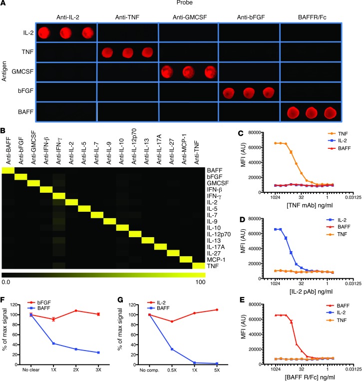
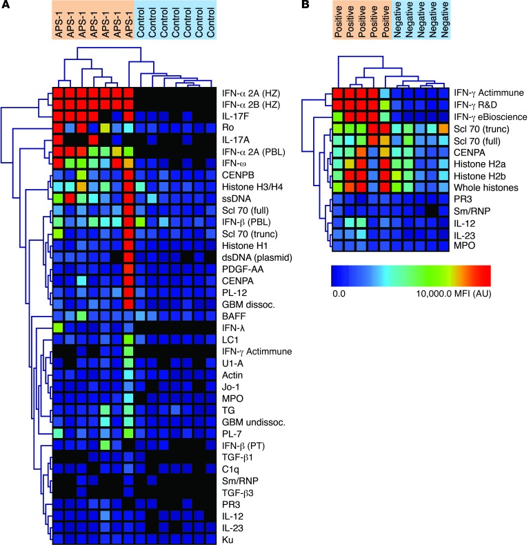
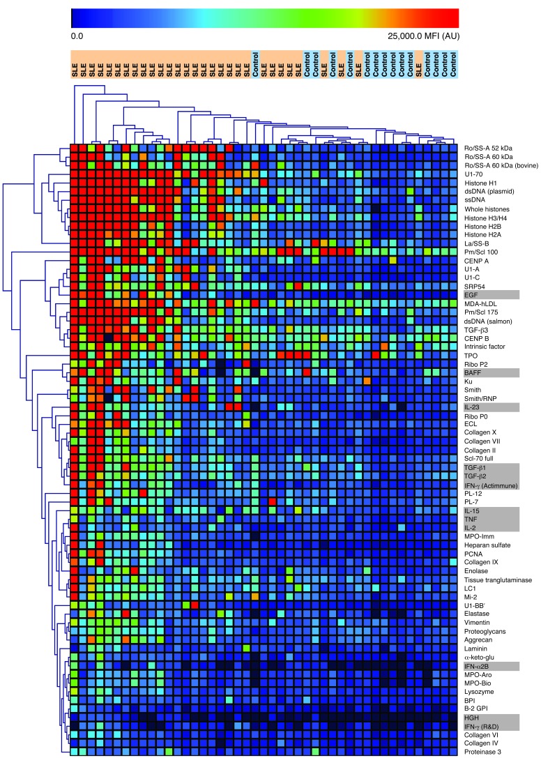
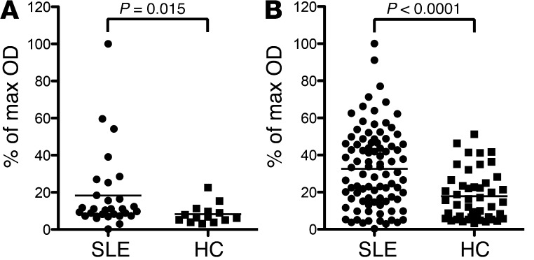
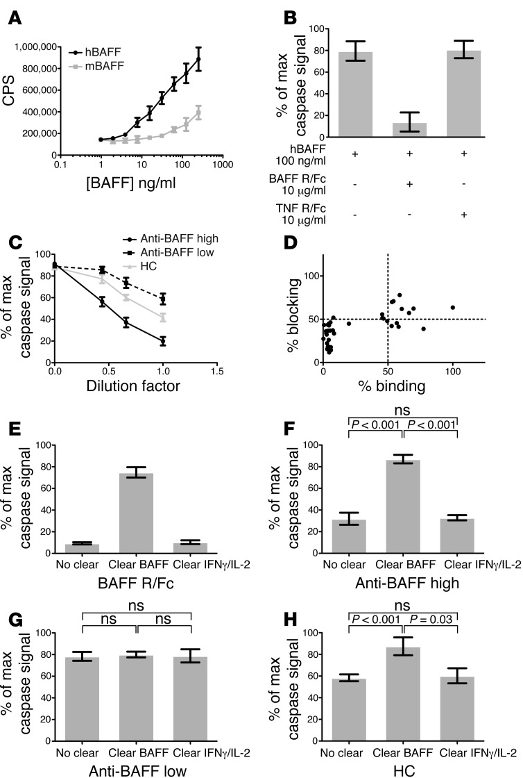
Autoantibodies against cytokines, chemokines, and growth factors inhibit normal immunity and are implicated in inflammatory autoimmune disease and diseases of immune deficiency. In an effort to evaluate serum from autoimmune and immunodeficient patients for Abs against cytokines, chemokines, and growth factors in a high-throughput and unbiased manner, we constructed a multiplex protein microarray for detection of serum factor-binding Abs and used the microarray to detect autoantibody targets in SLE. We designed a nitrocellulose-surface microarray containing human cytokines, chemokines, and other circulating proteins and demonstrated that the array permitted specific detection of serum factor-binding probes. We used the arrays to detect previously described autoantibodies against cytokines in samples from individuals with autoimmune polyendocrine syndrome type 1 and chronic mycobacterial infection. Serum profiling from individuals with SLE revealed that among several targets, elevated IgG autoantibody reactivity to B cell-activating factor (BAFF) was associated with SLE compared with control samples. BAFF reactivity correlated with the severity of disease-associated features, including IFN-α-driven SLE pathology. Our results showed that serum factor protein microarrays facilitate detection of autoantibody reactivity to serum factors in human samples and that BAFF-reactive autoantibodies may be associated with an elevated inflammatory disease state within the spectrum of SLE.


Figures

Figure 1
Figure 1. Validation of serum factor array specificity and dynamic range.
(A) Qualitative visualization of array results showing the specificity of serum factor–targeted probes for cognate antigen printed in triplicate. 5 separate arrays were probed with Abs against IL-2, TNF, GMCSF, bFGF, and BAFF R/Fc (see Methods and Supplemental Table 1). Reactivity was detected using Cy5-conjugated secondary Abs. Fluorescent images of indicated array features are shown for each probing condition, with each antigen shown in triplicate. (B) Heatmap showing proportion of array-wide maximum MFI at the indicated antigens for arrays probed separately with 16 different Abs. (CE) Range of detection for reagents (C) TNF mAb, (D) IL-2 pAb, and (E) BAFF R/Fc binding to cognate targets on serum factor microarrays. Data are mean ± SEM. (F) Array reactivity of a mixture of Abs targeting bFGF and BAFF, either uncleared (No clear) or subjected to 3 rounds of preclearing (1×, 2×, 3×) with BAFF-conjugated beads. Data (mean ± SEM) are shown as percent maximum MFI signal (relative to uncleared). (G) Array reactivity of a mixture of Abs targeting IL-2 and BAFF, either without competition (No comp) or incubated with 0.5:1, 1:1, or 5:1 molar ratios of unconjugated BAFF cytokine/Ab (0.5×, 1.0×, 5.0×, respectively). Data (mean ± SEM) are shown as percent maximum MFI signal (relative to no competition).
Figure 2
Figure 2. Serum factor array analysis of samples from individuals with APS-1 and nontuberculous DMAC.
(A and B) Heatmaps displaying serum IgG reactivity significantly associated with (A) APS-1 disease state versus HCs or (B) anti–IFN-γ–positive nontuberculous DMAC samples versus anti–IFN-γ–negative mycobacterial infection controls, as assessed by the SAM algorithm by 10,000 permutations of the data (q < 0.001).
Figure 3
Figure 3. Serum factor array reactivity in a cohort of individuals with SLE.
Heatmap displaying serum IgG reactivity significantly associated with SLE disease state versus HCs, as assessed by the SAM algorithm by 10,000 permutations of the data (q < 0.001). Serum factor antigens are shaded gray.
Figure 4
Figure 4. Detection of BAFF reactivity in 2 independent SLE cohorts by ELISA.
(A and B) BAFF reactivity detected in (A) ABCoN and (B) SCIDRR SLE sample cohorts and associated controls (see Supplemental Table 2). Values are presented as a percentage of the sample in each cohort with the highest OD (assigned as 100%). P values (2-tailed) represent significance of BAFF reactivity association with SLE versus HC in each cohort, assessed using Mann-Whitney U test.
Figure 5
Figure 5. Detection of BAFF blocking activity in SLE and HC samples using FL-17 cells.
(A) Caspase luminescence signal in wells containing FL-17 cells incubated with a serial dilution of soluble recombinant human and mouse BAFF (hBAFF and mBAFF, respectively), showing a sensitivity range of 1.0–250.0 ng/ml. CPS, counts per second. (B) Luminescent signal of FL-17 cells incubated with recombinant human BAFF in the presence of no inhibitor, BAFF R/Fc, or TNF R/Fc. (C) Inhibition of luminescence signal of FL-17 cells incubated with 100 ng/ml recombinant human BAFF in the presence of a serial dilution of IgG purified from individual anti-BAFF–high (n = 15), anti-BAFF–low (n = 15), and HC (n = 10) serum samples. P < 0.001 for all between-group comparisons, 2-way ANOVA and Tukey correction for multiple comparisons. (AC) Data represent mean ± SEM of ≥4 replicate wells. (D) Relationship between BAFF binding and BAFF blocking level in each of 40 SCIDRR cohort samples in the FL-17 assay. Blocking percentage was calculated as average CPS per sample normalized to maximum signal (no sample condition). (EH) Caspase luminescence signal in FL-17 cells incubated with 100 ng/ml BAFF in the presence of (E) 10 μg/ml BAFF R/Fc, or serum IgG derived from (F) 6 anti-BAFF–high, (G) 4 anti-BAFF–low, and (H) 4 HC samples from the SCIDRR cohort, with or without preclearing with sepharose beads conjugated to BAFF or IFN-γ/IL-2. Data are mean ± SEM. P values (2-tailed) were calculated by Mann-Whitney U test.

Comment in

  • BAFF-ling autoantibodies.
    Sarantopoulos S, Su MA. Sarantopoulos S, et al. J Clin Invest. 2013 Dec;123(12):5006-8. doi: 10.1172/JCI73166. Epub 2013 Nov 25. J Clin Invest. 2013. PMID: 24270413 Free PMC article.

References

    1. Morimoto AM, et al. Association of endogenous anti-interferon-α autoantibodies with decreased interferon-pathway and disease activity in patients with systemic lupus erythematosus. Arthritis Rheum. 2011;63(8):2407–2415. doi: 10.1002/art.30399. - DOI - PMC - PubMed
    1. Uchida K, et al. GM-CSF autoantibodies and neutrophil dysfunction in pulmonary alveolar proteinosis. N Engl J Med. 2007;356(6):567–579. doi: 10.1056/NEJMoa062505. - DOI - PubMed
    1. Doffinger R, et al. Autoantibodies to interferon-gamma in a patient with selective susceptibility to mycobacterial infection and organ-specific autoimmunity. Clin Infect Dis. 2004;38(1):e10–e14. doi: 10.1086/380453. - DOI - PubMed
    1. Hoflich C, et al. Naturally occurring anti-IFN-gamma autoantibody and severe infections with Mycobacterium cheloneae and Burkholderia cocovenenans. Blood. 2004;103(2):673–675. doi: 10.1182/blood-2003-04-1065. - DOI - PubMed
    1. Patel SY, et al. Anti-IFN-γ autoantibodies in disseminated nontuberculous mycobacterial infections. J Immunol. 2005;175(7):4769–4776. - PubMed

Publication types

MeSH terms

Associated data