. 2014 Feb;17(2):215-22.
doi: 10.1038/nn.3607. Epub 2013 Dec 22.

Distribution, recognition and regulation of non-CpG methylation in the adult mammalian brain

Affiliations

Distribution, recognition and regulation of non-CpG methylation in the adult mammalian brain

Junjie U Guo et al. Nat Neurosci. 2014 Feb.

Abstract

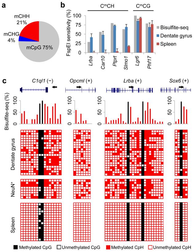
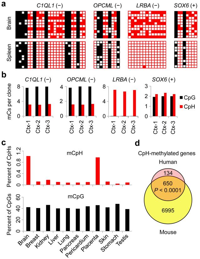
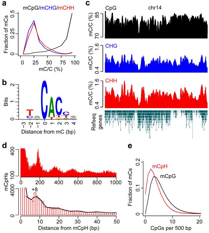
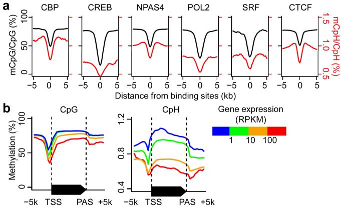
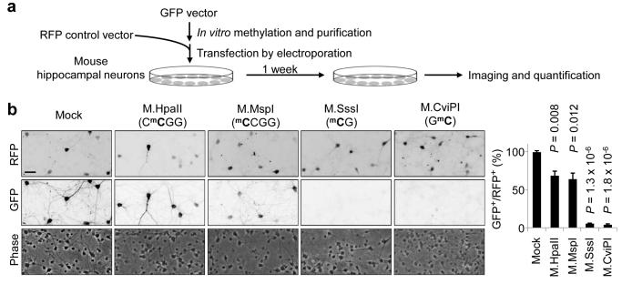
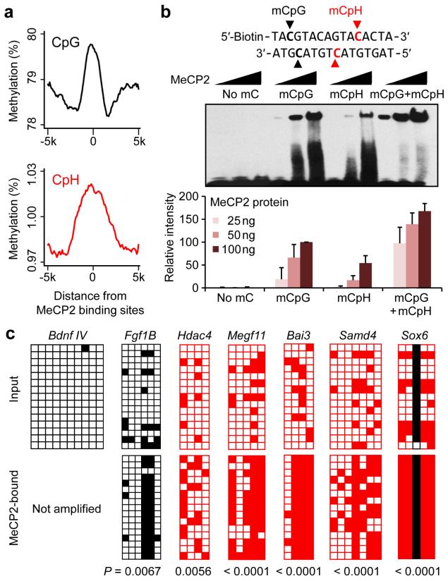
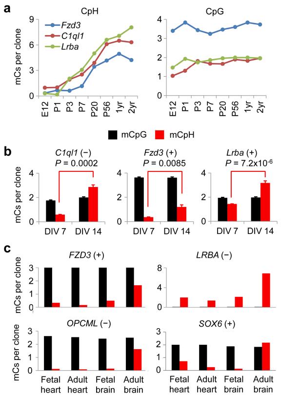
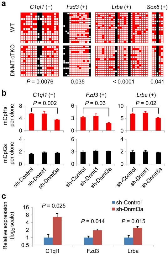
DNA methylation has critical roles in the nervous system and has been traditionally considered to be restricted to CpG dinucleotides in metazoan genomes. Here we show that the single base-resolution DNA methylome from adult mouse dentate neurons consists of both CpG (~75%) and CpH (~25%) methylation (H = A/C/T). Neuronal CpH methylation is conserved in human brains, enriched in regions of low CpG density, depleted at protein-DNA interaction sites and anticorrelated with gene expression. Functionally, both methylated CpGs (mCpGs) and mCpHs can repress transcription in vitro and are recognized by methyl-CpG binding protein 2 (MeCP2) in neurons in vivo. Unlike most CpG methylation, CpH methylation is established de novo during neuronal maturation and requires DNA methyltransferase 3A (DNMT3A) for active maintenance in postmitotic neurons. These characteristics of CpH methylation suggest that a substantially expanded proportion of the neuronal genome is under cytosine methylation regulation and provide a new foundation for understanding the role of this key epigenetic modification in the nervous system.


Figures

Figure 1
Figure 1. Pervasive CpH methylation in the in vivo DNA methylome of adult dentate granule neurons
(a) Composition of all mC loci in the genome of adult mouse dentate granule neurons. (b) Genomic DNA samples from adult dentate granule neurons were digested at methylated CmC motifs by FspEI. Sensitivity to FspEI digestion was measured by qPCR using primers flanking the predicted digestion sites (Supplementary Table 1a). Values represent mean ± s.e.m. (n = 3). Bisulfite-Seq results for each region are indicated by grey bars. (c) Four CpH-methylated loci were further examined by Sanger bisulfite sequencing in independent adult mouse dentate gyrus and spleen samples and FACS-sorted NeuN+ neuronal nuclei. Each row represents one DNA clone. Each column represents one mC site. Unmethylated and methylated cytosines are represented by open and filled boxes, respectively. CpG and CpH methylation are color-coded (black, CpG; red, CpH). Corresponding Bisulfite-Seq results are shown on the top panel.
Figure 2
Figure 2. Conserved CpH methylation in orthogolous regions of the human brain DNA
(a) Sanger bisulfite sequencing results of orthologous regions in the adult human brain and spleen genomic DNA. (b) Consistent levels of CpH methylation in multiple adult human cortical genomic DNA samples (Ctx1–3). (c) Reduced representation bisulfite sequencing (RRBS) data generated by the ENCODE project were analyzed using the mitochondrial CpH methylation rate (~1%) as the background probability. Percentage of mCpG/mCpH was corrected by the FDR estimated by a binomial distribution. (d) Quantification of numbers of mCpHs for each of the 15,417 one-to-one orthologous gene pairs between mouse and human (Ensembl annotations). A gene was considered CpH-methylated if two or more CpHs were ≥25% methylated (P value is indicated, χ2 test)
Figure 3
Figure 3. Genomic features of the neuronal CpH methylation
(a) Distributions of methylation levels of mCpG (black), mCHG (blue) and mCHH (red) in the genome of adult mouse dentate granule neurons. (b) Motif analysis of all mCpHs, with mCs being in position 0. (c) A chromosome-wide view of three types of mCs on chromosome 14 (chr14). Methylation levels were moving-averaged by 100 kb windows. Refseq transcript annotation is shown at the bottom. (d) Spacing analysis of adjacent mCpHs. The cubic spline smoothing curve is shown. Please see comparison between adult dentate granule neurons and embryonic stem cells in Supplementary Fig. 5. (e) Relationship between mCpG/mCpH occurrence and their local CpG densities.
Figure 4
Figure 4. Relationship between CpH methylation and protein-DNA interaction or gene expression in vivo
(a) Averaged methylation levels at previously profiled protein-DNA interaction sites. Data references for protein-DNA interactions are listed in Supplementary Table 2. CBP: CREB-binding protein; CREB: cAMP responsive element-binding protein; NPAS4: neuronal PAS domain protein 4; POL2: RNA polymerase II; SRF: serum response element-binding transcription factor; CTCF: CCCTC-binding factor. (b) Averaged methylation levels across all annotated genes, stratified by their mRNA levels in the adult mouse dentate gyrus. Both CpG and CpH methylation were anti-correlated with expression levels of associated genes. TSS: transcription start site; PAS: poly(A) site; RPKM: reads per kilobase of exon model per million mapped reads. Please see anti-correlation between CpG-far CH methylation in regulatory regions and gene expression in Supplementary Fig. 7.
Figure 5
Figure 5. Repression of reporter gene expression by CpH methylation in neurons
(a) A schematic diagram of experimental design of the in vitro methylated plasmid reporter assay. (b) Representative phase-contrast and immunofluorescent images of cultured hippocampal neurons co-transfected with an unmethylated RFP plasmid and GFP plasmids with different methylation patterns (left). Scale bar: 50 μm. Also shown is quantification of percentages of GFP+RFP+ neurons among all RFP+ neurons (right). Values represent mean ± s.e.m. (n = 3;P values are indicated for each condition; one-way ANOVA and Tukey’s test).
Figure 6
Figure 6. Recognition of CpH methylation by MeCP2 in vitro and in vivo
(a) Neuronal CpG (top) and CpH (bottom) methylation levels were averaged around MeCP2-bound regions previously determined using whole-mouse brains. (b) EMSA analysis using four oligo probes methylated at specific positions indicated in bold (top). Quantification of MeCP2-bound oligos is also shown (bottom). Values represent mean ± s.e.m. (n = 3). See Supplementary Fig. 9 for results of MBD2b. (c) MeCP2 ChIP-bisulfite-sequencing analysis of unmethylated, CpG-methylated/CpH-unmethylated, and CpH-methylated regions in the adult mouse hippocampus. The levels of CpH methylation in the latter regions were significantly higher in ChIP samples than in the input DNA from adult mouse hippocampus (P values are indicated; Fisher’s exact test).
Figure 7
Figure 7. Establishment of CpH methylation during neuronal maturation
(a) Progression of CpH (left) and CpG (right) methylation levels in the mouse brain at eight developmental time points. Values represent means (n = 3). (b) CpG and CpH methylation in mouse hippocampal neuronal cultures. Methylation levels were measured after 7 or 14 days in vitro (DIV). Values represent mean + s.e.m. (n = 3; P values are indicated; Student’s t test). (c) CpG and CpH methylation in fetal and adult human tissues.
Figure 8
Figure 8. Neuronal CpH methylation is actively maintained by DNMT3A and regulates endogenous gene expression in vivo
(a) CpH methylation levels were significantly decreased in postnatal neuron-specific DNMT1/3a/3b triple knockout (cTKO; CamKIIa-Cre). Samples from adult hippocampus were examined (P values are indicated; Fisher’s exact test). (b) Requirement of DNMT3A for maintenance of CpH methylation in the adult dentate gyrus. AAVs expressing different shRNAs were stereotaxically injected into the adult mouse dentate gyrus. DNA methylation was measured one week later from micro-dissected dentate gyri. Values represent mean ± s.e.m. (n = 3; P values are indicated; Student’s t-test). (c) mRNA expression of CpH-methylated genes following DNMT3A knock-down. Values represent mean ± s.e.m. (n = 3; P values are indicated; Student’s t-test). See also Supplementary Fig. 11 for results on unmethylated and CpG-methylated/CpH-unmethylated genes.

References

    1. Borrelli E, Nestler EJ, Allis CD, Sassone-Corsi P. Decoding the epigenetic language of neuronal plasticity. Neuron. 2008;60:961–74. - PMC - PubMed
    1. Feng J, Fan G. The role of DNA methylation in the central nervous system and neuropsychiatric disorders. Int Rev Neurobiol. 2009;89:67–84. - PubMed
    1. Day JJ, Sweatt JD. DNA methylation and memory formation. Nat Neurosci. 2010;13:1299–1440. - PMC - PubMed
    1. Zhang TY, Meaney MJ. Epigenetics and the environmental regulation of the genome and its function. Annu Rev Psychol. 2010;61:439–66. C1-3. - PubMed
    1. Ma DK, et al. Epigenetic choreographers of neurogenesis in the adult mammalian brain. Nat Neurosci. 2010;13:1338–1344. - PMC - PubMed

Publication types

MeSH terms

Associated data