. 2014 Sep 30:5:5055.
doi: 10.1038/ncomms6055.

A random six-phase switch regulates pneumococcal virulence via global epigenetic changes

Affiliations

A random six-phase switch regulates pneumococcal virulence via global epigenetic changes

Ana Sousa Manso et al. Nat Commun. .

Abstract

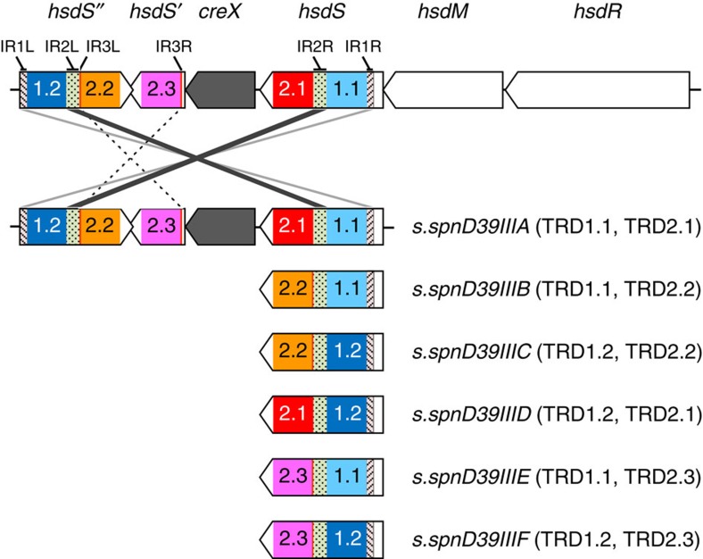
Streptococcus pneumoniae (the pneumococcus) is the world's foremost bacterial pathogen in both morbidity and mortality. Switching between phenotypic forms (or 'phases') that favour asymptomatic carriage or invasive disease was first reported in 1933. Here, we show that the underlying mechanism for such phase variation consists of genetic rearrangements in a Type I restriction-modification system (SpnD39III). The rearrangements generate six alternative specificities with distinct methylation patterns, as defined by single-molecule, real-time (SMRT) methylomics. The SpnD39III variants have distinct gene expression profiles. We demonstrate distinct virulence in experimental infection and in vivo selection for switching between SpnD39III variants. SpnD39III is ubiquitous in pneumococci, indicating an essential role in its biology. Future studies must recognize the potential for switching between these heretofore undetectable, differentiated pneumococcal subpopulations in vitro and in vivo. Similar systems exist in other bacterial genera, indicating the potential for broad exploitation of epigenetic gene regulation.


Conflict of interest statement

M.B., T.A.C. and J.K. are full-time employees at Pacific Biosciences, a company commercializing single-molecule, real-time nucleic acid sequencing technologies. The remaining authors declare no competing financial interests.

Figures

Figure 1
Figure 1. Schematic map of the SpnD39III locus and of the six alternative hsdS genes.
In strain D39, the Type I RM system SpnD39III locus (upper row from right to left) includes hsdR (locus_tag SPD_0455; GenBank protein database accession code ABJ53942; REBASE SpnD39ORF782P), hsdM (SPD_0454; ABJ54850; M.SpnD39ORF454P) and hsdS (SPD_0453; ABJ53819; S3.SpnD39ORF454P), a Cre recombinase (SPD_0452; ABJ54057), a truncated hsdS′ gene with only one variable domain (SPD_0450; ABJ55250; S2.SpnD39ORF454P) and a further truncated hsdS′′ gene with two variable domains (SPD_0451; ABJ54176; S1.SpnD39ORF454P). Three series of inverted repeats (IR1 to IR3) allow for recombination. These rearrangements, exemplified by crosses between the IRs, allow formation of six different variant hsdS alleles encoding different HsdS proteins SpnD39IIIA-F. Inverted repeats are of 85 (IR1, striped), 333 (IR2, dotted) and 15 bp (IR3). The six recombinant s.spnD39IIIA-F alleles (Genbank nucleotide database accession codes KJ955483, KJ955484, KJ955485, KJ955486, KJ398403 and KJ398404) are shown below with their TRDs numbered according to Table 1.
Figure 2
Figure 2. SpnD39III restriction.
(ah) Restriction by SpnD39III was demonstrated by transformation of differently methylated pDP28 plasmids into strains expressing only one SpnD39III variant. Restriction was evident in strains expressing variant SpnD39IIIB (b) and SpnD39IIIC (c), which have non-co-directional target sites on pDP28 (Supplementary Fig. 4), whereas restriction was not evident in strains expressing variants SpnD39IIIA (a) or SpnD39IIID (d), which have co-directional target sites on pDP28. Graphs indicate s.d. of three independent replicates. Statistically significant differences found using an analysis of variance Tukey test are shown. When utilizing recombinant pDP28 derivatives with two non-co-directional target sites for SpnD39IIIA (e) or SpnD39IIID (h) restriction could also be observed for these sites, whereas no restriction was observed when transforming a recombinant pDP28 with only one SpnD39IIIB site (f) or two co-directional SpnD39IIIC sites (g). ***P<0.001.
Figure 3
Figure 3. In vitro phenotypes of SpnD39IIIA-F variant strains.
(a) Phagocytosis of pneumococci by RAW 264.7 macrophages. Mean values of three independent replicates and s.d. are shown. (b) LuxS expression was assayed by quantitative western blot and the mean of quadruplicate samples are shown (in case of SpnD39E-F including two technical replicates). (c) The production of the type 2 polysaccharide capsule was quantified using an uronic acid assay and the mean and s.d. of quadruplicate samples are shown (in case of SpnD39E-F including two technical replicates). Statistical analysis of in vitro experiments (ac) was performed using one-way analysis of variance and a Tukey post-comparison test. *P<0.05, **P<0.01, ***P<0.001.
Figure 4
Figure 4. In vivo phenotypes of SpnD39IIIA-F variant strains.
D39 wt or derivatives expressing only a locked SpnD39IIIA to SpnD39IIIF variant were used in these experiments. Carriage experiments were performed by intranasal inoculation of 5 × 10⁴ pneumococci into BALB/c mice, which are resistant to invasive infection. (ac) The extent of carriage was evaluated by nasal lavage at day 1 (a), day 3 (b) and day 7 (c). (d,e) In the invasive disease model, susceptible CD1 mice received an intravenous challenge of 1 × 10⁵ pneumococci, and blood samples were taken at 4 and 30 h post challenge. Statistical significance of differing bacterial load in D39 wt or SpnD39III variant infected mice was calculated on log-transformed data using the unpaired (two-tailed) t-test. (f) Blood samples plated directly onto catalase agar plates were examined and the percentage of opaque colonies observed for each SpnD39III variant strain. (g,h) Allele quantification in D39 wt was performed on DNA extracted from the nasal lavages of the carriage experiment (see also Supplementary Table 5) or on DNA extracted from colonies grown from blood samples in the invasive disease experiment and SpnD39III variant percentages for each are represented respectively in g (inoculum black, 1 day grey, 3 days white, and 7 days striped) and h (inoculum black, 4 h grey and 30 h white). *P<0.05, **P<0.01, ***P<0.001.

Comment in

  • R-M systems go on the offensive.
    Lees J, Gladstone RA. Lees J, et al. Nat Rev Microbiol. 2015 Mar;13(3):131. doi: 10.1038/nrmicro3435. Epub 2015 Feb 2. Nat Rev Microbiol. 2015. PMID: 25639682 No abstract available.

References

    1. Donati C. et al. Structure and dynamics of the pan-genome of Streptococcus pneumoniae and closely related species. Genome Biol. 11, R107 (2010). - PMC - PubMed
    1. Webster L. T. & Clow A. D. Intranasal virulence of pneumococci for mice. J. Exp. Med. 58, 465–483 (1933). - PMC - PubMed
    1. Weiser J. N., Austrian R., Sreenivasan P. K. & Masure H. R. Phase variation in pneumococcal opacity: relationship between colonial morphology and nasopharyngeal colonization. Infect. Immun. 62, 2582–2589 (1994). - PMC - PubMed
    1. Srikhanta Y. N., Fox K. L. & Jennings M. P. The phasevarion: phase variation of type III DNA methyltransferases controls coordinated switching in multiple genes. Nat. Rev. Microbiol. 8, 196–206 (2010). - PubMed
    1. Loenen W. A., Dryden D. T., Raleigh E. A. & Wilson G. G. Type I restriction enzymes and their relatives. Nucleic Acids Res. 42, 20–44 (2014). - PMC - PubMed

Publication types

Substances