. 2014 Nov 25:5:5488.
doi: 10.1038/ncomms6488.

Exosomes secreted by nematode parasites transfer small RNAs to mammalian cells and modulate innate immunity

Affiliations

Exosomes secreted by nematode parasites transfer small RNAs to mammalian cells and modulate innate immunity

Amy H Buck et al. Nat Commun. .

Erratum in

Abstract

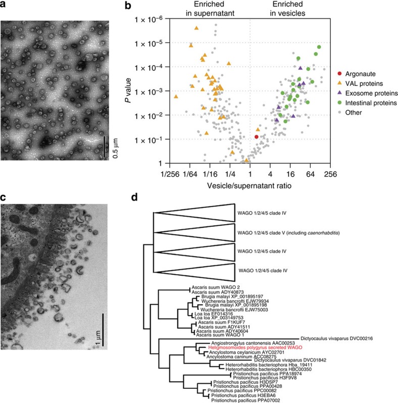
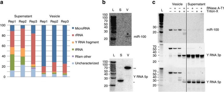
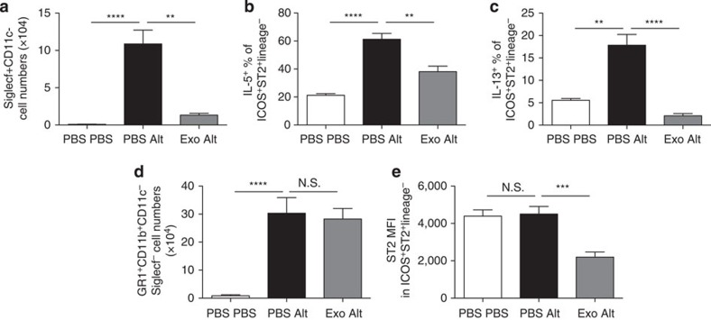
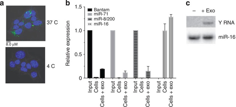
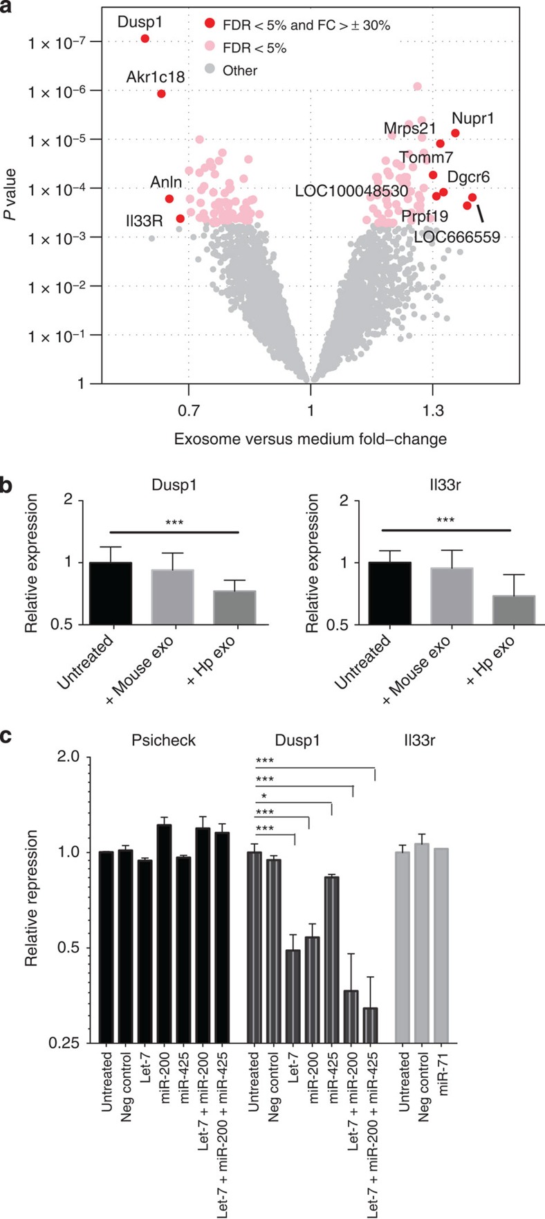
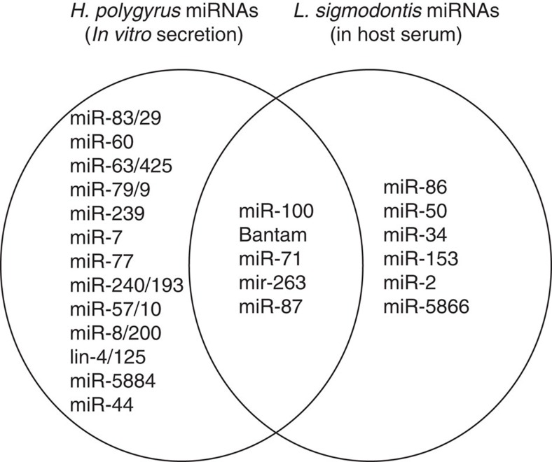
In mammalian systems RNA can move between cells via vesicles. Here we demonstrate that the gastrointestinal nematode Heligmosomoides polygyrus, which infects mice, secretes vesicles containing microRNAs (miRNAs) and Y RNAs as well as a nematode Argonaute protein. These vesicles are of intestinal origin and are enriched for homologues of mammalian exosome proteins. Administration of the nematode exosomes to mice suppresses Type 2 innate responses and eosinophilia induced by the allergen Alternaria. Microarray analysis of mouse cells incubated with nematode exosomes in vitro identifies Il33r and Dusp1 as suppressed genes, and Dusp1 can be repressed by nematode miRNAs based on a reporter assay. We further identify miRNAs from the filarial nematode Litomosoides sigmodontis in the serum of infected mice, suggesting that miRNA secretion into host tissues is conserved among parasitic nematodes. These results reveal exosomes as another mechanism by which helminths manipulate their hosts and provide a mechanistic framework for RNA transfer between animal species.


Figures

Figure 1
Figure 1. H. polygyrus secretory products contain miRNAs and Y RNAs.
(a) Size distribution of 3′-end labelled (pCp) total RNA extracted from the life stages (1 μg total RNA) or secretion product of H. polygyrus (total RNA from equivalent of 15 μg protein secretion product). (b) Proportion of H. polygyrus small RNA biotypes (<30 nt) identified in sequencing libraries from adult worms and the secretion product. (c) Predicted secondary structures of the two families of Y RNA identified in H. polygyrus secretion products based on RNAfold, the conserved UUAUC motif is noted in blue.
Figure 2
Figure 2. Many secreted nematode miRNAs have identical seed sites to mouse miRNAs.
(a) Temporal expression of highly abundant miRNAs (>10,000 reads per million in at least one of the libraries) across the life stages. Nematode and mouse names are listed according to identical seed sites and miRNAs of high abundance in the secretion product are coloured according to their conservation level: Eumetazoa (red), Bilateria (blue), Protostomia (green), Nematoda (orange). (b) Sequence alignment of abundant secreted parasite miRNAs that contain identical seed sites between mouse and H. polygyrus; all families shown are of common ancestry apart from miR-425/63.
Figure 3
Figure 3. H. polygyrus secretes exosomes of intestinal origin that contain a WAGO protein.
(a) TEM of purified ultracentrifugation pellet (100 μg ml−1 total protein) from H. polygyrus secretion product, scale indicates 0.5 μm. (b) Scatter plot of proteins enriched in ultracentrifugation pellet or supernatant based on LC-MS/MS, n=3, using P<0.05 (one-way ANOVA) and FC >1.5 as cutoffs. Noted in the legend are homologues of intestinal nematode proteins (green), mammalian exosome proteins (purple), Venom Allergen-Like (VAL) proteins (orange) and an Argonaute protein (red). (c) TEM of adult worm intestine noting vesicles of comparable size to exosomes, scale indicates 1.0 μm. (d) Phylogenetic relationship of the secreted Argonaute protein identified in H. polygyrus secretion product in relation to other nematode Argonautes. The analysis was performed on the same data set described in ref. with the addition of the H. polygyrus-secreted argonaute sequence, using the same method (Bayesian analysis using MrBayes v3.2).
Figure 4
Figure 4. Secreted miRNAs are protected from degradation through encapsulation within exosomes.
(a) Classification of H. polygyrus small RNAs in the secretion product following ultracentrifugation. (b) Northern blot analysis of RNA extracted from ultracentrifuge pellet or supernatant (from equivalent 10 μg protein) using probes complementary to H. polygyrus miR-100 or the 5′ arm of nematode Y RNA; * indicates the processed Y RNA and ** indicates the full length Y RNA. (c) Northern blot of RNA extracted from the pelleted secretion product following RNase treatment (0.5 Unit RNace-IT, 1 h at 37 °C) in the presence or absence of 0.05% Triton-X-100.
Figure 5
Figure 5. H. polygyrus exosomes suppress a Type 2 innate immune response in vivo.
H. polygyrus exosomes (10 μg) were administered intranasally to BALB/c mice 24 h before administration of 50 μg Alternaria extract and a further 5 μg exosomes, or controls that received PBS. (a) Siglecf+CD11c eosinophils in the bronchoalveolar lavage; (b) IL-5 and (c) IL-13 expression in PMA/ionomycin-stimulated lineage-negative, ICOS+ST2+ group 2 innate lymphoid cells in digested lung tissue were measured 24 h after Alternaria extract administration; (d) Gr1+CD11b+ neutrophils in the same lavage samples; (e) the mean fluorescence intensity (MFI) of ST2 (IL33R) staining in ILCs from each group of mice. Data are representative of two independent experiments, n=4–6 per group; error bars are mean±s.e.m. Data analysed by ANOVA and Tukey’s post test, ****P<0.0001, ***P<0.001, **P<0.01, *P<0.05.
Figure 6
Figure 6. H. polygyrus exosomes and RNAs are internalized by mouse cells.
(a) Confocal analysis of murine epithelial cells incubated for 1 h with PKH67-labelled H. polygyrus exosomes at 37 and 4 °C, scale indicates 8.0 μM. (b) Relative expression of parasite-derived miRNAs in murine epithelial cells at 20 h post incubation with 5 μg H. polygyrus exosomes following PBS washes. Signal observed in untreated host cells represents the background detection of the probe; for parasite-derived miRNA, the data are normalized to the input detection level of miRNAs in 5 μg of exosomes, whereas miR-16 levels in exosome-treated cells are normalized to untreated cells. (c) Northern blot analysis of RNA extracted from murine epithelial cells following 20 h incubation with H. polygyrus exosomes (5 μg total protein) compared with untreated cells following PBS washes, using a probe against the loop of the nematode Y RNA or mouse miR-16.
Figure 7
Figure 7. Mouse Il33r and Dusp1 are suppressed by H. polygyrus exosomes and the secreted miRNA repress target sites in Dusp1.
(a) Volcano plot of mouse genes up- or downregulated upon incubation with H. polygyrus-derived exosomes; red=FDR P<0.05 and FC>30%. (b) Levels of Dusp1 and Il1rl1 in mouse epithelial cells (5 × 10⁴) following 48 h treatment with 5 μg H. polygyrus exosomes or MODE-K-derived exosomes, n=8, error bars are mean±s.e.m. Data analysed by ANOVA and Tukey’s post test, *P<0.05, **P<0.01, ***P<0.005, ****P<0.001. (c) Repression of Psicheck reporter vector containing Dusp1 or Il1rl1 3′UTRs fused to Renilla luciferase by co-transfection with individual or pooled synthetic H. polygyrus miRNAs (50 nM), data represent renilla/luciferase ratios, normalized to the values obtained for untreated samples; n=3, ***P<0.005, *P<0.05.
Figure 8
Figure 8. Venn Diagram of overlap in miRNAs identified in H. polygyrus secretion product or serum of mice infected with L. sigmodontis.
The H. polygyrus miRNAs for comparison are taken from Supplementary Table 1 (top 20 most abundant in at least one platform). The miRNAs that are perfectly conserved between nematodes and mice are excluded, since the origin in serum cannot be deduced.

References

    1. Blaxter M. L. et al. A molecular evolutionary framework for the phylum Nematoda. Nature 392, 71–75 (1998). - PubMed
    1. Finney C. A., Taylor M. D., Wilson M. S. & Maizels R. M. Expansion and activation of CD4(+)CD25(+) regulatory T cells in Heligmosomoides polygyrus infection. Eur. J. Immunol. 37, 1874–1886 (2007). - PMC - PubMed
    1. Mohrs K., Harris D. P., Lund F. E. & Mohrs M. Systemic dissemination and persistence of Th2 and type 2 cells in response to infection with a strictly enteric nematode parasite. J. Immunol. 175, 5306–5313 (2005). - PubMed
    1. Rausch S. et al. Functional analysis of effector and regulatory T cells in a parasitic nematode infection. Infect. Immun. 76, 1908–1919 (2008). - PMC - PubMed
    1. McSorley H. J. & Maizels R. M. Helminth infections and host immune regulation. Clin. Microbiol. Rev. 25, 585–608 (2012). - PMC - PubMed

Publication types

MeSH terms

Associated data