. 2014 Jul 29:5:4510.
doi: 10.1038/ncomms5510.

Functionally diverse dendritic mRNAs rapidly associate with ribosomes following a novel experience

Affiliations

Functionally diverse dendritic mRNAs rapidly associate with ribosomes following a novel experience

Joshua A Ainsley et al. Nat Commun. .

Abstract

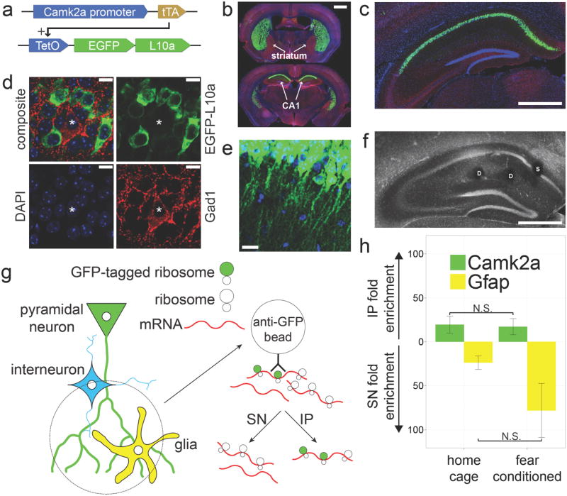
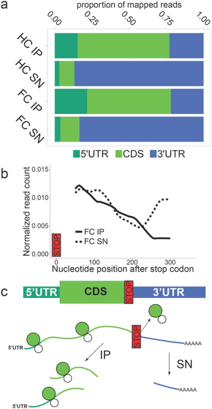
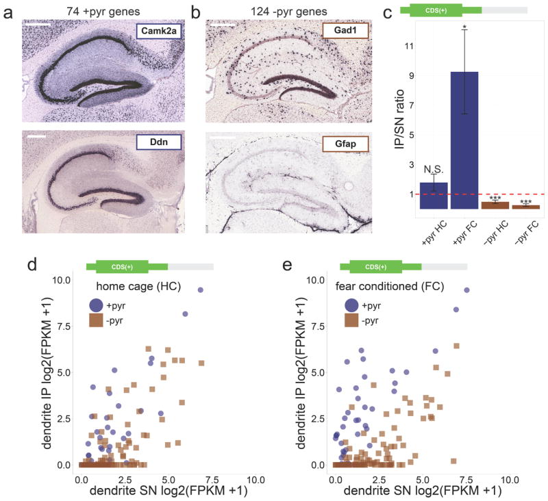
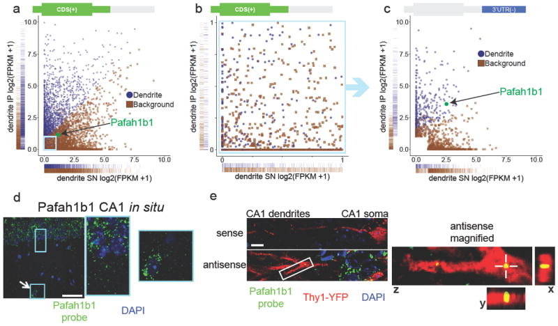
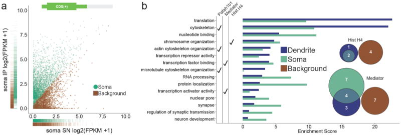
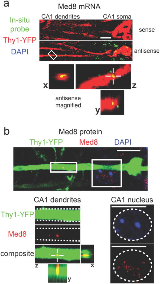
The subcellular localization and translation of messenger RNA (mRNA) supports functional differentiation between cellular compartments. In neuronal dendrites, local translation of mRNA provides a rapid and specific mechanism for synaptic plasticity and memory formation, and might be involved in the pathophysiology of certain brain disorders. Despite the importance of dendritic mRNA translation, little is known about which mRNAs can be translated in dendrites in vivo and when their translation occurs. Here we collect ribosome-bound mRNA from the dendrites of CA1 pyramidal neurons in the adult mouse hippocampus. We find that dendritic mRNA rapidly associates with ribosomes following a novel experience consisting of a contextual fear conditioning trial. High throughput RNA sequencing followed by machine learning classification reveals an unexpected breadth of ribosome-bound dendritic mRNAs, including mRNAs expected to be entirely somatic. Our findings are in agreement with a mechanism of synaptic plasticity that engages the acute local translation of functionally diverse dendritic mRNAs.


Conflict of interest statement

Competing financial interests

The authors declare no competing financial interests.

Figures

Figure 1
Figure 1. A novel method for collecting in vivo dendritic mRNA
(a) Camk2a-tTA, TetO-EGFP-L10a double transgenic mice were generated for cell type specific expression of EGFP-L10a. (b) EGFP-L10a expression is high in the striatum and dorsal CA1 (Green = EGFP-L10a, Blue = DAPI, Red = Gad1). Scale bar = 1mm (c) Within the hippocampus the expression of EGFP-L10a is limited to the CA1 region. Scale bar = 0.5mm (d) Within the CA1 region the expression of EGFP-L10a is restricted to excitatory pyramidal neurons (Green = EGFP-L10a, Blue = DAPI, Red = Gad1, asterisk indicates Gad1 positive cell). Scale bars = 10μm (e) EGFP-L10a is present in the dendrites of CA1 pyramidal neurons. Scale bar = 20μm (f) Representative example of the location of somatic (S) and dendritic (D) punches in the CA1. Scale bar = 0.5mm (g)Diagram showing the approach used for collecting ribosome-bound mRNA from in vivo dendrites. The immunoprecipitate (IP) will contain mRNA bound to ribosomes in CA1 pyramidal neuron dendrites while the supernatant (SN) will contain mRNA from other sources. (h) qPCR analysis of dendritic mRNA samples from home cage (n=6) and contextual fear conditioned (n=5) mice confirms the expected IP enrichment of Camk2a and SN enrichment of the astrocyte-specific gene Gfap. Error bars represent SEM.
Figure 2
Figure 2. RNA-Seq analysis confirms the ribosome-bound status of immunoprecipitated mRNA
(a) Dendritic immunoprecipitate (IP)samples had a smaller proportion of RNA-Seq reads mapping to the 3’UTR as compared to dendritic supernatant (SN) samples (HC: home cage, FC:contextual fear conditioning, UTR: untranslated region, CDS: coding sequence). (b) Depletion of 3’UTR reads in the dendritic IP started around 200 nucleotides after the stop codon as indicated by a decline in read coverage in the IP and a concomitant increase in read coverage in the SN. Read coverage was calculated for a set of genes that within the CA1 are only expressed in pyramidal neurons (+pyr genes, see Methods for details). (c)Diagram explaining how the depletion of 3’UTR reads in the IP confirms the expected ribosome-bound status of immunoprecipitated mRNA.As ribosomes bind to the 5’UTR and CDS of transcripts, random fragmenting of RNA would cause distal portions of the 3’UTR to remain in the SN.
Figure 3
Figure 3. Fear conditioning increases ribosome binding to dendritic mRNA
(a) Examples of +pyr control genes with exclusive expression only in CA1 pyramidal cells. Scale bar = 0.4mm (b) Examples of −pyr control genes with expression only outside of CA1 pyramidal cells ((a) and (b): images from Allen Mouse Brain Atlas). Scale bar = 0.4mm (c)In agreement with the enrichment of dendritic mRNA in the IP of dendritic punches, +pyr genes were on average enriched in the IP (IP/SN ratio > 1), while −pyr genes were on average enriched in the SN (IP/SN ratio < 1; CDS(+) FPKM values calculated from RNA-Seq data were used to calculate IP/SN ratios; red dashed line indicates IP/SN = 1; error bars represent SEM; Wilcoxon signed rank tests vs. IP/SN = 1 were performed for each group: N.S. = not significant, * = p < 0.05, *** = p < 0.0001). (d)Limited enrichment of dendritic mRNA in the home cage IP as indicated by a partial overlap between +pyr and −pyr genes in a scatterplot of IP and SN expression values (CDS(+) log2(FPKM+1) values calculated from the home cage dendritic RNA-Seq data were used to obtain expression values). (e)Fear conditioning increased enrichment of dendritic mRNA as indicated by increased separation between +pyr and −pyr genes (scatterplot generated similar to (d) except contextual fear conditioning dendritic RNA-Seq data were used). The increased enrichment of dendritic mRNA indicates increased ribosome binding of dendritic mRNA as a result of fear conditioning.
Figure 4
Figure 4. Machine learning prediction of dendritically localized mRNAs
(a) Classification of dendritic mRNAs by machine learning analysis of contextual fear conditioning dendritic RNA-Seq data (blue = dendritic, brown = background). One-dimensional density (rug) plots on the x and y axes show that mRNAs classified as dendritic have in general higher IP FPKMCDS(+) values and lower SN FPKMCDS(+)values when compared to mRNAs classified as background. One of the exceptions, Pafah1b1, is highlighted in green. Pafah1b1 is classified as dendritic despite having IP and SN FPKMCDS(+) values more similar to mRNAs classified as background. The light blue box indicates the area magnified in Fig. 4b. (b) Dendritic classification of mRNAs with low FPKMCDS(+)values (both IP and SN < 1) is not based on FPKMCDS(+) values as indicated by the intermixing of mRNAs classified as dendritic and background. (c) The same mRNAs as shown in Fig. 4b, this time plotted using the FPKM3’UTR(-) values. Pafah1b1 (green) is an example of an mRNA classified as dendritic mainly based on the FPKM3’UTR(-) values. Also see Supplementary Fig. 3e-i. (d) Pafah1b1 in situ using a FISH probe shows Pafah1b1 expression in the soma of pyramidal neurons (rectangular box) and an interneuron (square box with arrow) in the CA1 region. Scale bar = 50μm. (e) In situ hybridization of Pafah1b1 mRNA in Thy1-YFP transgenic tissue shows punctate labeling within dendrites, which is absent when using a sense probe (green = Pafah1b1 probe, red = Thy1-YFP, blue = DAPI). Scale bar = 10μm.On the right is a magnified view of the area indicated by the white box showing a Pafah1b1 mRNA puncta located within a YFP labeled dendrite (crosshair in z view marks same puncta shown in x and y views).
Figure 5
Figure 5. Gene ontology enrichment analysis reveals unexpected classes of ribosome-bound dendritic transcripts
(a) Classification of somatic mRNAs by machine learning analysis of contextual fear conditioning somatic RNA-Seq data (green = somatic, brown = background). (b) Gene ontology (GO) enrichment analysis of dendritic and somatic mRNAs.In agreement with previous studies , , dendritic mRNAs were highly enriched in translation and cytoskeleton GO categories. Unexpected GO categories with dendritic mRNA enrichment included chromosome organization and transcription factor binding. GO categories that include the Pafah1b1 gene, Mediator genes, and H4 histone genes are indicated. Venn diagram insets show the number of unique histone H4 and Mediator mRNAs that were detected in the RNA-Seq data as well as their classifications. Also see Supplementary Fig.4.
Figure 6
Figure 6. Dendritic localization of mRNA and protein encoded by the chromatin-associated gene Hist1h4j
(a) In situ hybridization for Hist1h4j shows puncta within YFP-labeled dendrites with the antisense probe, but not with the sense probe (green = Hist1h4j probe, red = YFP, blue = DAPI). The bottom panels show magnified views of the area indicated by the white box. Views from all three planes show colocalization between Hist1h4j mRNA and the YFP-labeled dendrite. (b)Histone H4 IHC results in dendritically-localized puncta (green = Thy1-YFP, red = Histone H4, blue = DAPI). Magnified dendritic and somatic areas are indicated with white boxes. Dashed white lines in the magnified dendritic image outline a single dendrite. Views from all three planes show colocalization between Histone H4 protein and the dendrite. A dashed white circle in the magnified somatic image outlines a nucleus with the expected presence of Histone H4 protein. Scale bars = 10μm.
Figure 7
Figure 7. Dendritic localization of mRNA and protein encoded by the transcription-associated gene Med8
(a) In situ hybridization for Med8 shows puncta within YFP-labeled dendrites with the antisense probe, but not with the sense probe (green = Med8 probe, red = YFP, blue = DAPI). The bottom panels show magnified views of the area indicated by the white box. Views from all three planes show colocalization between Med8 mRNA and the YFP- labeled dendrite. (b) Med8 IHC results in dendritically-localized puncta (green = Thy1-YFP, red = Med8, blue = DAPI). Magnified dendritic and somatic areas are indicated with white boxes.Dashed white lines in the magnified dendritic image outline a single dendrite. Views from all three planes show colocalization between Med8 protein and the dendrite. A dashed white circle in the magnified somatic image outlines a nucleus with the expected presence of Med8 protein. Scale bars = 10μm.

References

    1. Bodian DA. Suggestive Relationship of Nerve Cell Rna with Specific Synaptic Sites. Proc Natl Acad Sci U S A. 1965;53:418–425. - PMC - PubMed
    1. Steward O, Levy WB. Preferential localization of polyribosomes under the base of dendritic spines in granule cells of the dentate gyrus. J Neurosci. 1982;2:284–291. - PMC - PubMed
    1. Ostroff LE, Fiala JC, Allwardt B, Harris KM. Polyribosomes redistribute from dendritic shafts into spines with enlarged synapses during LTP in developing rat hippocampal slices. Neuron. 2002;35:535–545. - PubMed
    1. Holt CE, Schuman EM. Perspective The Central Dogma Decentralized : New Perspectives on RNA Function and Local Translation in Neurons. Neuron. 2013;80:648–57. - PMC - PubMed
    1. Miller S, et al. Disruption of Dendritic Translation of CaMKII[alpha] Impairs Stabilization of Synaptic Plasticity and Memory Consolidation. Neuron. 2002;36:507–519. - PubMed

Publication types

MeSH terms

Associated data