. 2015 Mar 6;10(3):e0119177.
doi: 10.1371/journal.pone.0119177. eCollection 2015.

Pregnane X receptor knockout mice display aging-dependent wearing of articular cartilage

Affiliations

Pregnane X receptor knockout mice display aging-dependent wearing of articular cartilage

Kotaro Azuma et al. PLoS One. .

Abstract

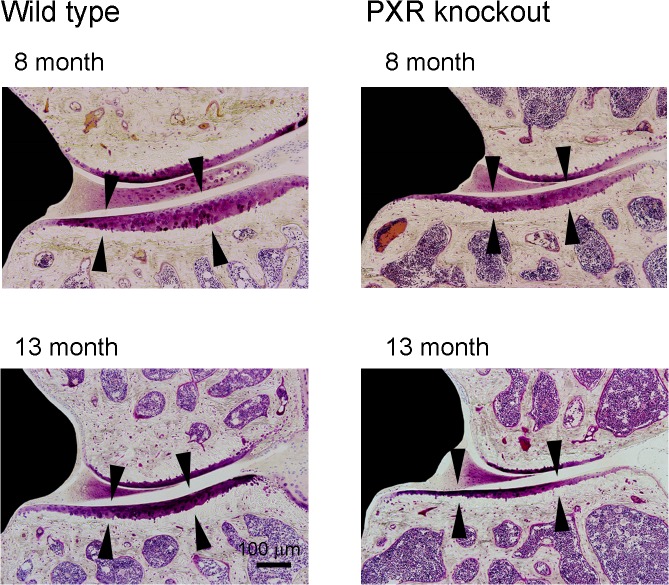
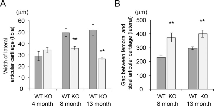
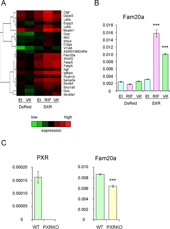
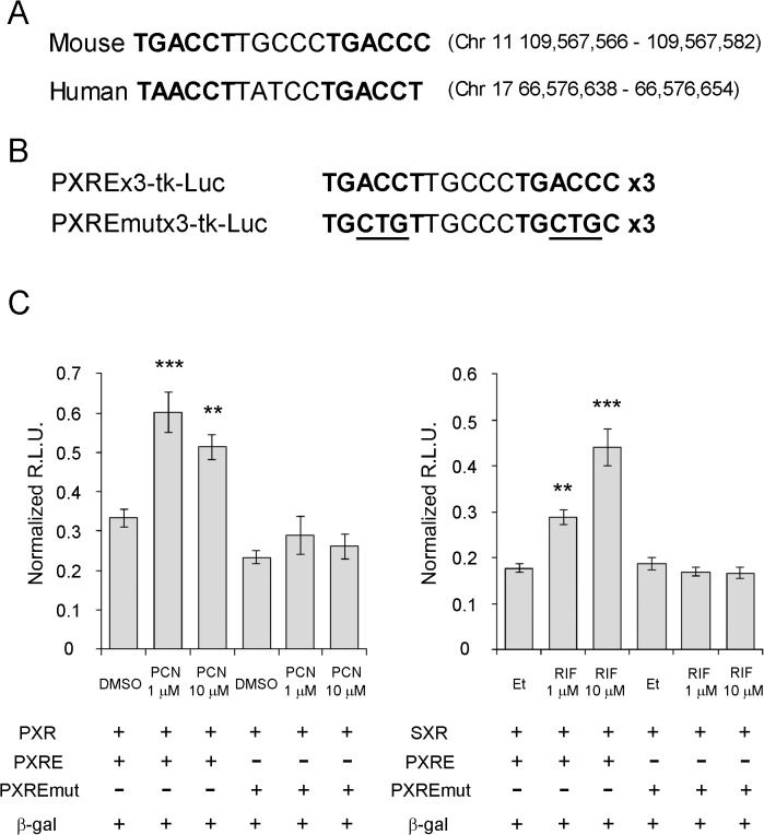
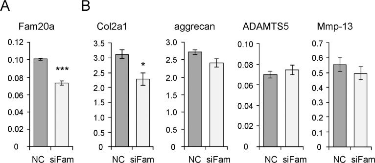
Steroid and xenobiotic receptor (SXR) and its murine ortholog, pregnane X receptor (PXR), are nuclear receptors that are expressed at high levels in the liver and the intestine where they function as xenobiotic sensors that induce expression of genes involved in detoxification and drug excretion. Recent evidence showed that SXR and PXR are also expressed in bone tissue where they mediate bone metabolism. Here we report that systemic deletion of PXR results in aging-dependent wearing of articular cartilage of knee joints. Histomorphometrical analysis showed remarkable reduction of width and an enlarged gap between femoral and tibial articular cartilage in PXR knockout mice. We hypothesized that genes induced by SXR in chondrocytes have a protective effect on articular cartilage and identified Fam20a (family with sequence similarity 20a) as an SXR-dependent gene induced by the known SXR ligands, rifampicin and vitamin K2. Lastly, we demonstrated the biological significance of Fam20a expression in chondrocytes by evaluating osteoarthritis-related gene expression of primary articular chondrocytes. Consistent with epidemiological findings, our results indicate that SXR/PXR protects against aging-dependent wearing of articular cartilage and that ligands for SXR/PXR have potential role in preventing osteoarthritis caused by aging.


Conflict of interest statement

Competing Interests: B. Blumberg is the holder of several US patents related to SXR which are controlled by the Salk Institute for Biological Studies and have been licensed to for-profit organizations. The authors confirm that this does not alter their adherence to all PLOS ONE policies on sharing data and materials.

Figures

Fig 1
Fig 1. Wearing of articular cartilage of the knee joint in PXR knockout mouse.
Representative microscopic image of articular cartilage of 8-month-old and 13-month-old wild-type and PXR knockout mice are shown. Arrowheads indicate lateral articular cartilage of the tibia.
Fig 2
Fig 2. Age-dependent wearing of articular cartilage of the knee joint.
(A) Width of lateral articular cartilage of the tibia in 4-month-old wild type (WT; n = 8) and PXR knockout (KO; n = 8) mice, 8 month-old wild type (WT; n = 6) and PXR knockout (KO; n = 6) mice, and 13 month-old wild type (WT; n = 5) and PXR knockout (KO; n = 4) mice is shown. (B) Gap between femoral and tibial articular cartilage of 8-month-old wild-type (WT; n = 6) and PXR knockout (KO; n = 6) mice and 13 month-old wild-type (WT; n = 5) and PXR knockout (KO; n = 4) mice are shown. **P < 0.01.
Fig 3
Fig 3. SXR-dependent induction of Fam20a by SXR ligands.
(A) ATDC5 cells were infected with adeno-SXR or adeno-DsRed and cultured in phenol red-free DMEM with charcoal/dextran-treated FCS (5%) containing rifampicin (RIF) (10 μM), vitamin K2 (VK) (10 μM), or ethanol (Et). Total RNA was extracted and gene expression was analyzed by microarray followed by hierarchical cluster analysis. A heat-map visualization of clusters including SXR-dependent ligand-induced genes is shown. Red grids indicate high expression and green grids indicate low expression. (B) SXR-dependent induction of Fam20a was validated by quantitative real-time PCR. Expression of GAPDH was used as an internal control. ***P < 0.001 in Dunnett’s test with adeno-SXR-infected ethanol-treated cells as a control. (C) Expression of Fam20a was decreased in primary articular chondrocytes derived from PXR knockout mice. Primary chondrocytes were purified from the femoral and knee joints of newborn PXR knockout mice and wild type mice. ***P < 0.001.
Fig 4
Fig 4. Consensus SXR/PXR responsive element motifs in the first introns of SXR and PXR genes.
(A) Consensus SXR/PXR responsive element motifs, variant direct repeat 5, were identified in the first intron of both murine PXR gene (chromosome 11) and human SXR gene (chromosome 17). The bold letters indicate consensus SXR/PXR binding motif. (B) Generation of reporter plasmid containing three copies of PXR responsive element (PXRE) and PXRE with mutation (PXREmut). Underlined letters indicate mutated nucleotides. (C) Cos7 cells were transfected with PXR or SXR expression vector and reporter plasmid containing murine PXR responsive element or mutated PXR responsive element, and β-galactosidase expression vector (β-gal). The cells were then treated with indicated concentrations of PXR agonist pregnane-16α-carbonitrile (PCN) or SXR agonist rifampicin (RIF) or vehicles: DMSO for PCN and ethanol (Et) for RIF. Data are shown as relative light units (R.L.U.) normalized by β-galactosidase activity. ** P < 0.01, ***P < 0.001 in Dunnett’s test with vehicle treated group as a control.
Fig 5
Fig 5. Physiological role of Fam20a in chondrocytes.
(A) Expression of Fam20a was knocked down using siRNA (siFam) in primary murine articular chondrocytes. The effect of the siRNA was evaluated by comparison with primary murine chondrocytes transfected with scrambled-sequence siRNA as a negative control (NC). Expression of Fam20a was analyzed by quantitative real-time PCR. Expression of GAPDH was used as an internal control. ***P < 0.001. (B) Analysis of osteoarthritis-related genes in Fam20a knocked-down cells. Expression of Col2a1 and aggrecan was measured as major proteins comprising the extracellular matrix of articular chondrocytes. Expression of ADAMTS5 and Mmp-13 was measured as major enzymes that degrade the extracellular matrix. Expression of GAPDH was used as an internal control. A significant decrease of Col2a1 expression was observed in Fam20a knocked-down chondrocytes. *P < 0.05.

References

    1. Blumberg B, Sabbagh W Jr, Juguilon H, Bolado J Jr, van Meter CM, Ong ES, et al. SXR, a novel steroid and xenobiotic-sensing nuclear receptor. Genes Dev. 1998;12: 3195–3205. - PMC - PubMed
    1. Synold TW, Dussault I, Forman BM. The orphan nuclear receptor SXR coordinately regulates drug metabolism and efflux. Nat Med. 2001;7: 584–590. - PubMed
    1. Zhou C, Verma S, Blumberg B. The steroid and xenobiotic receptor (SXR), beyond xenobiotic metabolism. Nucl Recept Signal. 2009;7: e001 10.1621/nrs.07001 - DOI - PMC - PubMed
    1. Azuma K, Ouchi Y, Inoue S. Vitamin K. Novel molecular mechanisms of action and its roles in osteoporosis. Geriatr Gerontol Int. 2014;14: 1–7. 10.1111/ggi.12236 - DOI - PubMed
    1. Tabb MM, Sun A, Zhou C, Grün F, Errandi J, Romero K, et al. Vitamin K2 regulation of bone homeostasis is mediated by the steroid and xenobiotic receptor SXR. J Biol Chem. 2003;278: 43919–43927. - PubMed

Publication types

MeSH terms

Associated data