. 2015 Apr 20;25(8):993-1004.
doi: 10.1016/j.cub.2015.02.019. Epub 2015 Mar 19.

Steroid Signaling Establishes a Female Metabolic State and Regulates SREBP to Control Oocyte Lipid Accumulation

Affiliations

Steroid Signaling Establishes a Female Metabolic State and Regulates SREBP to Control Oocyte Lipid Accumulation

Matthew H Sieber et al. Curr Biol. .

Abstract

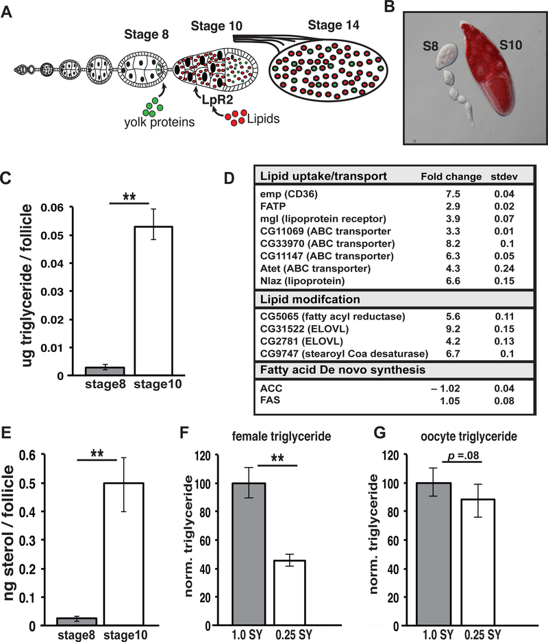
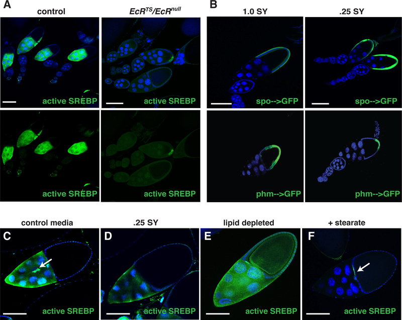
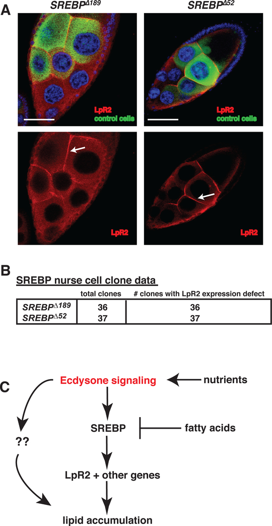
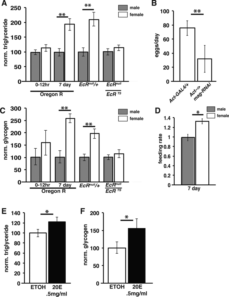
Disruptions in energy homeostasis severely affect reproduction in many organisms and are linked to several reproductive disorders in humans. As a result, understanding the mechanisms that control nutrient accumulation in the oocyte will provide valuable insights into the links between metabolic disease and reproductive dysfunction. We show that the steroid hormone ecdysone functions in Drosophila to control lipid metabolism and support oocyte production. First, local EcR-mediated signaling induces a stage-specific accumulation of lipids in stage-10 oocytes. EcR induces lipid accumulation by promoting the activation of the lipogenic transcription factor SREBP and by controlling the expression of the low-density lipoprotein (LDL) receptor homolog, LpR2. Second, global signaling via the ecdysone receptor, EcR, establishes a female metabolic state and promotes whole-body triglyceride and glycogen storage at high levels. EcR acts in the CNS to mediate these effects, in part by promoting higher levels of feeding in females. Thus, ecdysone functions at two levels to support reproduction: first by inducing lipid accumulation in the late stages of oocyte development and second by providing a signal that coordinates lipid metabolism in the germline with whole-animal lipid homeostasis. Ecdysone regulation allows females to assess the demands of oogenesis and alter their behavior and metabolic state to support the biosynthetic requirements of oocyte production.


Conflict of interest statement

Conflict of interest: The authors declare no conflict of interest.

Figures

Figure 1.
Figure 1.. Triglycerides and sterols accumulate during stage 10 of oogenesis
(A) Stages of Drosophila oogenesis showing germline lipid accumulation during stage 10. (B) Ovariole stained with Oil Red O (red). Triglyceride (C) and sterol (E) levels showing increases between stage 8 and stage 10 follicles. (D) RNA expression fold change between stage 8 and stage 10 of the indicated lipid metabolic genes as determined by RNAseq. (F, G) Whole female or stage 14 oocyte triglyceride levels are compared from animals fed on 0.25 SY or 1.0 SY food. TAG levels were normalized to total protein and are displayed as normalized to a wild-type level of 100% . Error bars represent 1xSD. **P<. 005
Figure 2.
Figure 2.. Ecdysone signaling is required for lipid accumulation during vitellogenesis
(A, B, C) Control and EcRts/EcRnull animals were dissected and ovaries were stained with DAPI (blue), anti-LpR2 antibodies (green), and Nile Red (red). Stained stage 10 egg chambers (follicles) from control n=208 and EcRts/EcRnull n=171 animals were scored (D) as having normal (A’), partial (B’) or absent (C’) LpR2 expression (Arrows point to LpR2 membrane staining). Error bars represent standard deviation. (E, F) Fold change in SCAP, SREBP and the indicated lipid metabolic gene mRNA levels as determined by RNA sequencing of EcRts/EcRnull compared to control stage 10 egg chambers. All error bars represent ± SD. Scale bars = 100um.
Figure 3.
Figure 3.. SREBP functions to promote lipid accumulation during vitellogenesis
Stage 10 follicles from control (A) and SREBP null mutant germ line clones (B, C), stained for DAPI (blue), clonal marker (anti-GFP: green), and Nile Red (red). Ovarioles from control (D) and nos–> SREBP-DBD animals stained with DAPI (blue) and Nile Red (red). Scale bars= 100um.
Figure 4.
Figure 4.. SREBP is regulated by Ecdysone signaling and dietary nutrients
(A) Follicles from control or EcRts/EcRnull animals carrying an SREBP-GFP reporter (green). (B) Follicles from phm–>GFP or spo–>GFP (green) animals fed either a control diet or a 0.25SY low calorie diet for 5 days. A stage 10 follicle from animals carrying an SREBP-GFP reporter (green) raised on a control diet (C), on a 0.25 SY low-calorie diet (D), on a lipid-depleted diet (E), or a 2% stearate supplemented diet (F). Arrows: border cells. Scale bars= 100um.
Figure 5.
Figure 5.. SREBP is required for LpR2 expression
(A) Stage 10 nurse cells with negatively marked SREBP mutant clones stained for DNA (DAPI: blue), clonal marker (GFP:green), and LpR2 (red). Arrows indicate lpr2 membrane staining. Scale bars = 50 um. (B) summary of nurse cell clones analyzed and their LpR2 membrane expression. (C) Proposed model of germline lipid accumulation.
Figure 6.
Figure 6.. Ecdysone signaling promotes triglyceride accumulation in adult females
Normalized adult triglyceride (A) and glycogen (C) levels from controls of the indicated genotypes and from EcRts/EcRnull mutant animals. (B) Egg laying in control and mag-RNAi expressing animals; n=40; error bar= 1xSD (D) Normalized feeding rate of mature adult males (= 1.0) and females via capillary assay. Triglyceride (E) and glycogen (F) in Oregon R males grown for 4 days on food supplemented with EtOH alone (=1.0) or with 0.5mg/ml 20-hydroxyecdysone . *p<0.005 **p<0.0001.
Figure 7.
Figure 7.. EcR functions in the central nervous system to promote feeding in females.
Triglyceride (A) and glycogen (B) in mature adult controls (UAS-EcR-DN/+) or in animals with CNS-driven (Elav-GAL4, GAL80ts) UAS-EcR-DN. (C) Egg laying/day by females from (UAS-EcR-DN/+) and (Elav-GAL4, GAL80ts) UAS-EcR-DN) animals, N=40; error bars= 1x SD. (D,E) Feeding rate via capillary assay for the indicated genotypes, normalized to control males = 1.0. (F) Model for the role of ecdysone in metabolic sexual dimorphism. *p<0.005 **p<0.0001.

References

    1. Shahjahan M, Kitahashi T, and Parhar IS (2014). Central Pathways Integrating Metabolism and Reproduction in Teleosts. Front Endocrinol (Lausanne) 5, 36. - PMC - PubMed
    1. Roland AV, and Moenter SM (2014). Reproductive neuroendocrine dysfunction in polycystic ovary syndrome: Insight from animal models. Front Neuroendocrinol. - PMC - PubMed
    1. Orbetzova MM, Kamenov ZA, Kolarov GB, Orbetzova VT, Genchev GD, Genov NS, and Zacharieva SZ (2003). Metabolic disturbances in women with polycystic ovary syndrome. Folia Med (Plovdiv) 45, 12–20. - PubMed
    1. Drummond-Barbosa D, and Spradling AC (2001). Stem cells and their progeny respond to nutritional changes during Drosophila oogenesis. Dev Biol 231, 265–278. - PubMed
    1. Yan J, Zhou B, Yang J, Tai P, Chen X, Zhang H, Zhang M, and Xia G (2008). Glucose can reverse the effects of acute fasting on mouse ovulation and oocyte maturation. Reprod Fertil Dev 20, 703–712. - PubMed

Publication types

MeSH terms

Associated data