. 2015 Oct 28;10(10):e0141836.
doi: 10.1371/journal.pone.0141836. eCollection 2015.

Transcriptomic Changes Due to Cytoplasmic TDP-43 Expression Reveal Dysregulation of Histone Transcripts and Nuclear Chromatin

Affiliations

Transcriptomic Changes Due to Cytoplasmic TDP-43 Expression Reveal Dysregulation of Histone Transcripts and Nuclear Chromatin

Alexandre Amlie-Wolf et al. PLoS One. .

Abstract

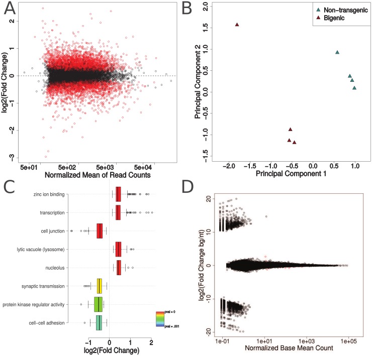
TAR DNA-binding protein 43 (TDP-43) is normally a nuclear RNA-binding protein that exhibits a range of functions including regulation of alternative splicing, RNA trafficking, and RNA stability. However, in amyotrophic lateral sclerosis (ALS) and frontotemporal lobar degeneration with TDP-43 inclusions (FTLD-TDP), TDP-43 is abnormally phosphorylated, ubiquitinated, and cleaved, and is mislocalized to the cytoplasm where it forms distinctive aggregates. We previously developed a mouse model expressing human TDP-43 with a mutation in its nuclear localization signal (ΔNLS-hTDP-43) so that the protein preferentially localizes to the cytoplasm. These mice did not exhibit a significant number of cytoplasmic aggregates, but did display dramatic changes in gene expression as measured by microarray, suggesting that cytoplasmic TDP-43 may be associated with a toxic gain-of-function. Here, we analyze new RNA-sequencing data from the ΔNLS-hTDP-43 mouse model, together with published RNA-sequencing data obtained previously from TDP-43 antisense oligonucleotide (ASO) knockdown mice to investigate further the dysregulation of gene expression in the ΔNLS model. This analysis reveals that the transcriptomic effects of the overexpression of the ΔNLS-hTDP-43 transgene are likely due to a gain of cytoplasmic function. Moreover, cytoplasmic TDP-43 expression alters transcripts that regulate chromatin assembly, the nucleolus, lysosomal function, and histone 3' untranslated region (UTR) processing. These transcriptomic alterations correlate with observed histologic abnormalities in heterochromatin structure and nuclear size in transgenic mouse and human brains.


Conflict of interest statement

Competing Interests: The authors have declared that no competing interests exist.

Figures

Fig 1
Fig 1. Transcriptomic analysis of ΔNLS-hTDP-43 expressing mice.
(A), Comparison of normalized mean of read counts with log2(fold change) shows no bias for highly expressed genes to be categorized as differentially expressed. 4,321 out of 10,601 genes meeting the minimum read count threshold were found to be significantly differentially expressed in ΔNLS-hTDP-43 expressing mice compared to nontransgenic mice (Benjamini-Hochberg corrected p-value < 0.05, red points). (B), Multidimensional scaling and principal component analysis shows distinct global transcriptomic profiles for ΔNLS-hTDP-43 bigenic (n = 4) and control nontransgenic (n = 4) mice. (C), Pathway analysis of significantly changing genes between bigenic and control mice reveals an upregulation in pathways predominantly involved in nuclear molecular processes and a downregulation in pathways relating to neuronal or glial function. (D), Splicing analysis reveals that only 23 out of 250,620 exons (red dots) are significantly differentially expressed with Benjamini-Hochberg correct p-values of less than 0.05.
Fig 2
Fig 2. Nuclear and cytosolic fractionation of ΔNLS-hTDP-43 expressing mice.
Subcellular fractionation followed by immunoblot protein analysis reveals a similar level of nuclear total (human and mouse, h+m) TDP-43 levels between bigenic (+/+) and control (+/- or -/-) mice, but an increase in cytosolic TDP-43 levels in the bigenic mice. HSP90 and histone H3 confirms separation of the cytosolic and nuclear fractions, respectively. n = 4 mice per category (4 bigenic, 2 monogenic, 2 non-transgenic).
Fig 3
Fig 3. Transcriptomic comparison between ΔNLS-hTDP-43 overexpression and TDP-43 ASO knockdown.
(A), Transcriptomic comparison of the 10,345 genes meeting the minimum read count in both experiments shows no correlation between ΔNLS-hTDP expression and TDP-43 ASO knockdown (Pearson r = 0.011). (B), Differential expression analysis reveals that 3,714 out of 11,916 genes meeting the minimum read count threshold were significantly differentially expressed (Benjamini-Hochberg corrected p-value < 0.05, red points) upon ASO knockdown, with no bias of read counts on significance. (C), Multidimensional scaling and principal component analysis shows distinct global transcriptomic profiles for TDP-43 ASO knockdown (n = 4) and control ASO (n = 4) mice. (D), Comparison of genes meeting the minimum read counts in both experiments shows that although the majority of genes are present in both experiments, those that are significantly differentially expressed show very little overlap between ΔNLS-hTDP expression and TDP-43 ASO knockdown. (E), Splicing analysis shows that 1,029 out of 250,620 exons are differentially expressed upon ASO-mediated TDP-43 knockdown. (F), Pathway analysis suggests that most of the transcriptomic effects due to ASO knockdown are associated with the wound response and neuronal death.
Fig 4
Fig 4. Histone transcript dysregulation in ΔNLS-hTDP-43 mice.
(A), Comparison of multimapped read fold change in canonical and variant histones shows a significant upregulation in canonical histones (fold change = 2.12, 2-sided t-test p = 0.046) and significant but slight downregulation in variant histones (fold change = 0.841, 2-sided t-test p = 0.017) in transgenic vs nontransgenic mice. (B), Analysis of log2(fold changes) of genes involved in histone 3’ UTR processing reveal an enrichment of significantly changing genes. (C), Analysis of TDP-43 CLIP binding on genes involved in histone 3’ UTR processing shows enrichment for CLIP binding. (D), RT-qPCR validation of histone 3’ UTR processing genes and canonical histones with an oligo-dT selection for polyadenylated RNA confirms the upregulation in aberrantly polyadenylated histone genes in the ΔNLS-TDP-43-expressing mice. (E), Similar RT-qPCR validation with random hexamer priming shows that non-polyadenylated canonical histones are not significantly upregulated in the bigenic mice. (F), Comparison of RNA-seq and RT-PCR analysis validates RNA-seq results, except for histone transcripts (white filled symbols) which revealed distinct results when priming with random hexamers versus oligo-dT’s. *p < 0.05, **p < 0.01, *** p < 0.001, **** p < 0.0001.
Fig 5
Fig 5. Altered nuclear morphology associated with cytoplasmic TDP-43 protein.
(A) Cresyl violet staining of bigenic mice brain sections reveals abnormal nucleolar and chromatin structure and nucleomegaly affecting hippocampal pyramidal (CA1) neurons, olfactory bulb granular neurons, and neocortical neurons. Cerebellar granular and Purkinje neurons, where the transgene is not expressed, show no nuclear morphologic change. (B) Representative confocal immunofluorescence image of human hippocampal dentate gyrus neuronal nuclei stained with DAPI (blue) with and without TDP-43 inclusions (red). (C) Quantification of average cross sectional nuclear area of neurons without TDP-43 inclusions (n = 254) and neurons with TDP-43 inclusions (n = 133) from 13 different FTLD-TDP cases. Median value of nuclear area for neurons without TDP-43 inclusions is 57.56 μm2 and is 75.66 μm2 for neurons with TDP-43 inclusions. Paired t-test reveals a significant difference in nuclear size (p < 0.0001).

References

    1. Ou S, Wu F, Harrich D, García-Martínez L, Gaynor R. Cloning and characterization of a novel cellular protein, TDP-43, that binds to human immunodeficiency virus type 1 TAR DNA sequence motifs. J Virol. 1995; - PMC - PubMed
    1. Buratti E, Baralle FE. Characterization and functional implications of the RNA binding properties of nuclear factor TDP-43, a novel splicing regulator of CFTR exon 9. J Biol Chem. 2001. September 28;276(39):36337–43. - PubMed
    1. Lagier-Tourenne C, Polymenidou M, Cleveland DW. TDP-43 and FUS/TLS: emerging roles in RNA processing and neurodegeneration. Hum Mol Genet. 2010. April 15;19(R1):R46–64. 10.1093/hmg/ddq137 - DOI - PMC - PubMed
    1. Chen-Plotkin AS, Lee VM-Y, Trojanowski JQ. TAR DNA-binding protein 43 in neurodegenerative disease. Nat Rev Neurol. 2010. March;6(4):211–20. 10.1038/nrneurol.2010.18 - DOI - PMC - PubMed
    1. Ling S-C, Polymenidou M, Cleveland DW. Converging mechanisms in ALS and FTD: disrupted RNA and protein homeostasis. Neuron. Elsevier Inc.; 2013. August 7;79(3):416–38. - PMC - PubMed

Publication types

MeSH terms

Associated data