

Cutting Edge: IL-36 Receptor Promotes Resolution of Intestinal Damage
- PMID: 26590314
- PMCID: PMC4684965
- DOI: 10.4049/jimmunol.1501312
Cutting Edge: IL-36 Receptor Promotes Resolution of Intestinal Damage
Abstract
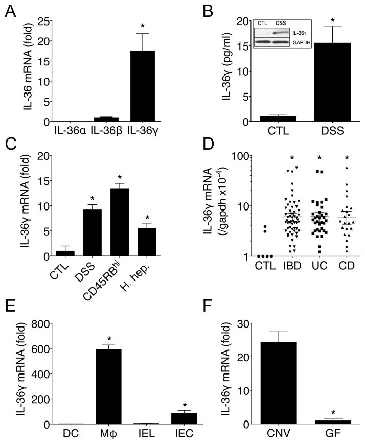
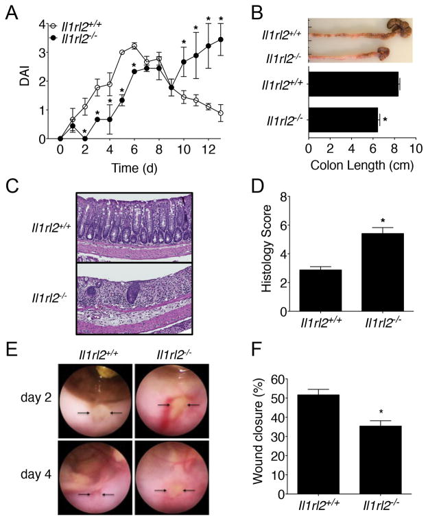
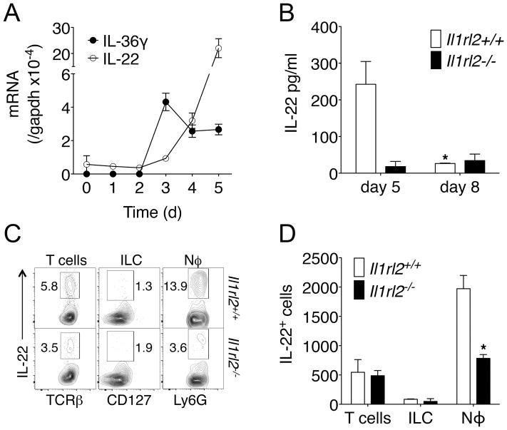
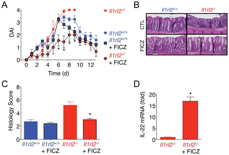
IL-1 family members are central mediators of host defense. In this article, we show that the novel IL-1 family member IL-36γ was expressed during experimental colitis and human inflammatory bowel disease. Germ-free mice failed to induce IL-36γ in response to dextran sodium sulfate (DSS)-induced damage, suggesting that gut microbiota are involved in its induction. Surprisingly, IL-36R-deficient (Il1rl2(-/-)) mice exhibited defective recovery following DSS-induced damage and impaired closure of colonic mucosal biopsy wounds, which coincided with impaired neutrophil accumulation in the wound bed. Failure of Il1rl2(-/-) mice to recover from DSS-induced damage was associated with a profound reduction in IL-22 expression, particularly by colonic neutrophils. Defective recovery of Il1rl2(-/-) mice could be rescued by an aryl hydrocarbon receptor agonist, which was sufficient to restore IL-22 expression and promote full recovery from DSS-induced damage. These findings implicate the IL-36/IL-36R axis in the resolution of intestinal mucosal wounds.
Copyright © 2015 by The American Association of Immunologists, Inc.
Figures
References
-
- Takagi H, Kanai T, Okazawa A, Kishi Y, Sato T, Takaishi H, Inoue N, Ogata H, Iwao Y, Hoshino K, Takeda K, Akira S, Watanabe M, Ishii H, Hibi T. Contrasting action of IL-12 and IL-18 in the development of dextran sodium sulphate colitis in mice. Scand J Gastroenterol. 2003;38:837–844. - PubMed
-
- Dupaul-Chicoine J, Yeretssian G, Doiron K, Bergstrom KS, McIntire CR, LeBlanc PM, Meunier C, Turbide C, Gros P, Beauchemin N, Vallance BA, Saleh M. Control of intestinal homeostasis, colitis, and colitis-associated colorectal cancer by the inflammatory caspases. Immunity. 2010;32:367–378. - PubMed
Publication types
MeSH terms
Substances
Associated data
- Actions
Grants and funding
- R01 DK079392/DK/NIDDK NIH HHS/United States
- R29 DK055679/DK/NIDDK NIH HHS/United States
- R01 DK097256/DK/NIDDK NIH HHS/United States
- T32 GM008169/GM/NIGMS NIH HHS/United States
- DK72564/DK/NIDDK NIH HHS/United States
- R01 DK072564/DK/NIDDK NIH HHS/United States
- P30 DK034933/DK/NIDDK NIH HHS/United States
- DK61739/DK/NIDDK NIH HHS/United States
- DK055679/DK/NIDDK NIH HHS/United States
- DK79392/DK/NIDDK NIH HHS/United States
- R01 DK055679/DK/NIDDK NIH HHS/United States
- DK097256/DK/NIDDK NIH HHS/United States
- DK059888/DK/NIDDK NIH HHS/United States
- R01 DK059888/DK/NIDDK NIH HHS/United States
LinkOut - more resources
Full Text Sources
Other Literature Sources
Molecular Biology Databases
