. 2024 Nov 30;15(1):10434.
doi: 10.1038/s41467-024-54764-3.

SARS-CoV-2 human challenge reveals biomarkers that discriminate early and late phases of respiratory viral infections

Affiliations

SARS-CoV-2 human challenge reveals biomarkers that discriminate early and late phases of respiratory viral infections

Joshua Rosenheim et al. Nat Commun. .

Abstract

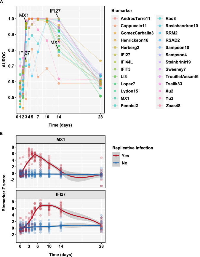
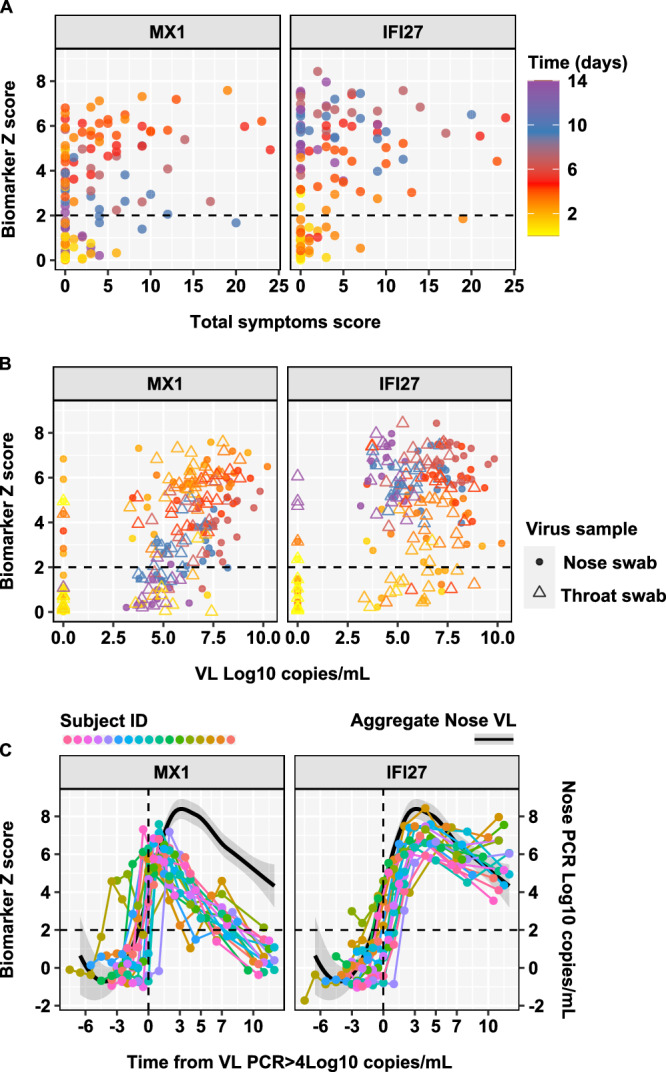
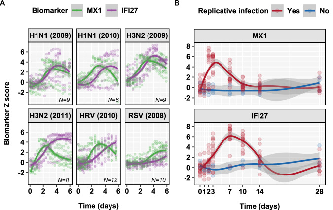
Blood transcriptional biomarkers of acute viral infections typically reflect type 1 interferon (IFN) signalling, but it is not known whether there are biological differences in their regulation that can be leveraged for distinct translational applications. We use high frequency sampling in the SARS-CoV-2 human challenge model to show induction of IFN-stimulated gene (ISG) expression with different temporal and cellular profiles. MX1 gene expression correlates with a rapid and transient wave of ISG expression across all cell types, which may precede PCR detection of replicative infection. Another ISG, IFI27, shows a delayed but sustained response restricted to myeloid cells, attributable to gene and cell-specific epigenetic regulation. These findings are reproducible in experimental and naturally acquired infections with influenza, respiratory syncytial virus and rhinovirus. Blood MX1 expression is superior to IFI27 expression for diagnosis of early infection, as a correlate of viral load and for discrimination of virus culture positivity. Therefore, MX1 expression offers potential to stratify patients for antiviral therapy or infection control interventions. Blood IFI27 expression is superior to MX1 expression for diagnostic accuracy across the time course of symptomatic infection and thereby, offers higher diagnostic yield for respiratory virus infections that incur a delay between transmission and testing.


Conflict of interest statement

Competing interests: The Authors declare the following competing interests: In the past 3 years, S.A.T. has received remuneration for scientific advisory board membership from Sanofi, GlaxoSmithKline, Foresite Labs and Qiagen. S.A.T. is a co-founder and holds equity in Transition Bio and Ensocell. From 8 January 2024, S.A.T. has been a part-time employee of GlaxoSmithKline. A.J.M., A.C., M.K., M.M. and A.B. are full time employees at hVIVO Services Ltd. No other authors report any competing interests.

Figures

Fig. 1
Fig. 1. SARS-CoV-2 PCR viral load in nose and throat swabs following virus challenge.
A Schematic overview of SARS-CoV-2 challenge of unvaccinated SARS-CoV-2 seronegative healthy adults showing time points for viral load measurements (nose and throat swabs), and host RNA sequencing (blood samples and nose swabs). Blue shading represents period of quarantine. B Quantitative viral load measurements by PCR from nose and (C) throat swabs per participant (rows) stratified by time point (columns) after virus challenge, and clustered (using complete linkage hierarchical clustering) into two groups of participants with (N = 17) and without (N = 16) evidence of replicative virus infection. Grey colour denotes unavailable data points.
Fig. 2
Fig. 2. Blood transcriptional discrimination of participants with and without replicative infection by time from SARS-CoV-2 challenge.
A Point estimates for area under the receiver operating characteristic curve (AUROC) stratified by blood transcriptional signature and time after virus challenge. B Individual (data points) and loess smoothed summary (line ±95% CI) for standardised blood transcript levels of MX1 and IFI27 in sequential time points after challenge, for participants with (N = 17) and without (N = 16) replicative viral infection.
Fig. 3
Fig. 3. Relationship between blood transcript levels of MX1 and IFI27 with symptoms and viral load by time from SARS-CoV-2 challenge.
Individual standardised blood transcript levels of MX1 and IFI27 against (A) symptom scores and (B) nose and throat viral loads in participants who developed replicative virus infection (N = 17), stratified by time after virus challenge, with dashed line to represent the threshold (Z > 2) for elevated transcript levels. C Individual standardised blood transcript levels of MX1 and IFI27 (connected data points, left axis) and loess smoothed summary for nose viral load (black line ± 95% CI, right axis) in participants who developed replicative virus infection (N = 17), by time from virus detection in nose swabs > 4 Log10 copies/mL.
Fig. 4
Fig. 4. Discrimination of virus culture positivity by blood transcript levels of MX1 and IFI27.
A Individual standardised blood transcript levels of MX1 and IFI27 at each time point for all individuals who develop replicative infection (N = 17), stratified by contemporary virus culture positivity in either nose or throat swabs, with dashed line to represent the threshold (Z > 2) for elevated transcript levels. B Area under the receiver operating characteristic curve (AUROC) discrimination of virus culture positivity by blood transcript levels of MX1 and IFI27 across all time points, showing AUROC point estimates and 95% confidence intervals.
Fig. 5
Fig. 5. Differential regulation of MX1 and IFI27 expression in blood.
Individual standardised blood transcriptional expression of a type 1 IFN stimulated gene signature (STAT1 module) by standardised blood transcriptional expression of (A) MX1 and (B) IFI27 at all time points in participants who developed replicative virus infection (N = 17), showing linear regression lines ±95% CI, 2-sided Spearmen correlation coefficients and p value. C Standardised blood transcriptional expression of MX1 and IFI27 stratified by cell type and time after virus challenge, in pooled data from participants who developed replicative virus infection (N = 6).
Fig. 6
Fig. 6. Generalisable differences in temporal profiles of blood MX1 and IFI27 expression in diverse respiratory virus challenges.
Individual (data points) and loess smoothed summaries (lines ±95% CI) for (A) standardised blood transcript levels of MX1 and IFI27 over the first 6 days after challenge in selected human respiratory virus challenge models (GSE73072) among participants who develop replicative infection, and (B) over 14 days in an H3N2 influenza human challenge model, among participants with (N = 16) and without (N = 3) replicative infection.
Fig. 7
Fig. 7. Differences in temporal profiles of blood MX1 and IFI27 expression in naturally acquired respiratory virus infections, and delayed responses in the nose to virus challenge.
A Discrimination between SARS-CoV-2 infected (N = 20) and uninfected (N = 26) household contacts of index cases with COVID-19 by blood transcript levels of MX1 and IFI27, at participant recruitment (study day 0) and 7 days later. Data points represent individual study participants, summarised by box and whisker plots showing median ±interquartile range ± 1.5 xIQR. Discrimination accuracy is shown as AUROC point estimate and 95% confidence intervals. B Individual standardised blood transcript levels of MX1 against IFI27 for sequential samples before infection (baseline, N = 128) and at time points indicated (day0, N = 103; day 2, N = 106, day 4, N = 100; day 6, N = 102) after presentation within 48 hours of symptoms onset among prospectively recruited participants with unselected respiratory virus infections. C Loess smoothed summary (line ±95% CI) for standardised transcript levels of MX1 and IFI27 in blood (N = 17) and nose samples (N = 5-13 for different time points) from participants who developed replicative virus infection by time after SARS-CoV-2 challenge, and in blood (N = 16) and nose samples (N = 12-13 for different time points) from participants who developed replicative virus infection by time after H3N2 influenza challenge.

References

    1. Gupta, R. K. & Noursadeghi, M. Toward a more generalizable blood RNA signature for bacterial and viral infections. Cell Rep. Med3, 100866 (2022). - PMC - PubMed
    1. Gupta, R. K. et al. Blood transcriptional biomarkers of acute viral infection for detection of pre-symptomatic SARS-CoV-2 infection: a nested, case-control diagnostic accuracy study. Lancet Microbe2, e508–e517 (2021). - PMC - PubMed
    1. Swadling, L. et al. Pre-existing polymerase-specific T cells expand in abortive seronegative SARS-CoV-2. Nature601, 110–117 (2022). - PMC - PubMed
    1. Killingley, B. et al. Safety, tolerability and viral kinetics during SARS-CoV-2 human challenge in young adults. Nat. Med28, 1031–1041 (2022). - PubMed
    1. Andres-Terre, M. et al. Integrated, multi-cohort analysis identifies conserved transcriptional signatures across multiple respiratory viruses. Immunity43, 1199–1211 (2015). - PMC - PubMed

Publication types

MeSH terms

Associated data