. 2015 Sep 8;12(10):1691-703.
doi: 10.1016/j.celrep.2015.07.064. Epub 2015 Aug 28.

Telomerase Is Essential for Zebrafish Heart Regeneration

Affiliations

Telomerase Is Essential for Zebrafish Heart Regeneration

Dorota Bednarek et al. Cell Rep. .

Abstract

After myocardial infarction in humans, lost cardiomyocytes are replaced by an irreversible fibrotic scar. In contrast, zebrafish hearts efficiently regenerate after injury. Complete regeneration of the zebrafish heart is driven by the strong proliferation response of its cardiomyocytes to injury. Here we show that, after cardiac injury in zebrafish, telomerase becomes hyperactivated, and telomeres elongate transiently, preceding a peak of cardiomyocyte proliferation and full organ recovery. Using a telomerase-mutant zebrafish model, we found that telomerase loss drastically decreases cardiomyocyte proliferation and fibrotic tissue regression after cryoinjury and that cardiac function does not recover. The impaired cardiomyocyte proliferation response is accompanied by the absence of cardiomyocytes with long telomeres and an increased proportion of cardiomyocytes showing DNA damage and senescence characteristics. These findings demonstrate the importance of telomerase function in heart regeneration and highlight the potential of telomerase therapy as a means of stimulating cell proliferation upon myocardial infarction.


Figures

Figure 1
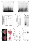
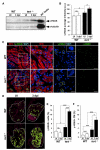
Figure 1. Heart Cryoinjury Augments Telomerase Activity and tert Expression Levels
(A) Representative telomeric repeat amplification protocol (TRAP) activity in uninjured (UI) hearts and hearts at 1, 3, 4, 7, and 30 days post cryoinjury (dpi) (n = 3/condition and time point). Positive control (C+), iPSCs; negative control (C−), tert−/− zebrafish heart; reaction specificity control, uninjured + RNase. LB, lysis buffer. (B and C) Telomerase TRAP activity assay in 6- and 10-month-old zebrafish hearts (n = 4/time point). Data are means ± SEM. ns, not significant (unpaired Student’s t test). (D) Zebrafish tert mRNA expression levels in homeostasis and during regeneration (3 dpi) (n = 4 hearts/condition). CPMs, counts per million. Values are means ± SEM. ***p < 0.001 (B–H adjusted p value). (E) Representative TRAP assay showing the lack of telomerase activity in tert−/− hearts (n = 3/genotype). (F) Lack of telomerase does not affect heart development and function. There is normal anatomy and function in tert−/− zebrafish hearts. Shown are whole-mount views of dissected adult WT and tert−/− uninjured zebrafish hearts (left) and Masson-Goldner trichrome-stained sagittal sections (center). Five animals were analyzed per genotype. V, ventricle; At, atrium; BA, bulbus arteriosus. Scale bars, 100 μm. (G and H) Echocardiographic evaluation of heart size (ventricular volume (VV) in diastole) (G) and cardiac function (ventricular FVS) (H) in uninjured WT and tert−/− animals. Values for both parameters did not differ between genotypes. Circles and squares show data for individual animals. Horizontal bars represent the mean (unpaired Student’s t test). A total of 17–20 animals were analyzed per genotype.
Figure 2
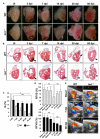
Figure 2. Heart Regeneration Is Inhibited Strongly in tert−/− Animals
(A) Whole-mount views of uninjured and cryoinjured WT and tert−/− zebrafish hearts dissected at the indicated times post-injury. Dotted lines outline the injured area. Asterisks mark the initial injury site. (B) Masson-Goldner trichrome staining of sagittal sections of uninjured WT and tert−/− hearts at the indicated days after cryoinjury. Dotted black lines outline the injured area. Arrows mark the initial injury site. Scale bars, 100 μm. (C) Size of the ventricle injury (injured area [IA]) on sagittal heart sections at the indicated times post-injury, presented as the percentage of the total ventricular area. Data are means of at least 4 sections/heart from 3-8 hearts/time point, with the exception of WT 30 dpi, where only one heart was analyzed. Data are means ± SEM. *p < 0.05 (Mann-Whitney test). (D) Relative FVS (RFVS) in WT and tert−/− zebrafish hearts under the basal condition (sham-operated animals) and at the indicated times post-injury. WT hearts recover ventricular function over time but tert−/− hearts do not. Data are means ± SEM of a pool of 10 animals/condition. **p < 0.01 (one-way ANOVA followed by Tukey’s honest significant difference test). (E) Representative Doppler echocardiography images of intracardial blood flow in WT and tert−/− hearts in the absence of injury and at the indicated times post-injury. Asterisks mark the initial injury site. See also Movies S1, S2, S3, S4, S5, and S6. See also Figures S1–S3, and Table S1.
Figure 3
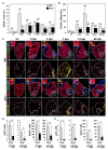
Figure 3. The Absence of Telomerase Severely Affects Cardiomyocyte Proliferation
(A and B) Cardiac cells positive for PCNA in UI WT and tert−/− zebrafish hearts and in hearts at the indicated days after cryoinjury. (A) PCNA+ total cardiac cells. (B) PCNA+ cardiomyocytes. Data are means ± SEM of the percentage of PCNA+ cells (A) or number of PCNA+ cardiomyocytes per cardiac ventricle section (at least 3 sections/animal from the indicated number of animals) (B). *p < 0.05 compared with WT samples (Mann-Whitney test). (C) WT and tert−/− heart sections immunostained with anti-MHC to mark cardiomyocytes (red) and anti-PCNA to mark cells in S phase (green). Nuclei were counterstained with DAPI (blue). The bottom rows show the location of PCNA+ cardiomyocytes during regeneration (yellow circles). The nuclear area is shown in magenta. Dotted lines outline the ventricle and injured area. Asterisks mark the initial injury site. Insets show high-magnification views of representative PCNA+ cardiomyocytes in the boxed areas. Scale bars, 100 μm (whole-heart views) and 10 μm (magnifications). (D and E) Quantification of pH3+ and BrdU-labeled cardiac cells (D) and cardiomyocytes (E) at 7 and 14 dpi. Data are means ± SEM. *p < 0.05, **p < 0.01 (unpaired Student’s t test). See also Figures S4 and S5.
Figure 4
Figure 4. Heart Cryoinjury Induces a Telomerase-Dependent Increase in Telomere Reserves
(A) Representative telomap images of WT and tert−/− heart, uninjured (UI) or fixed at different days postinjury [dpi]. Nuclei are assigned a four-color code according to their average telomere fluorescence in a.u. The cells with the longest telomeres are visualized in red, and the cells with the shortest telomeres are presented in green. Dotted lines mark the injured area. The second and fourth rows show higher magnifications of the boxed areas highlighted in the entire heart section images. Scale bars, 100 μm and 10 μm for magnification images. (B) Telomere fluorescence frequency histograms of cardiac WT and tert−/− cells in uninjured hearts and hearts at 3, 7, 14, 30, and 60 days after cryoinjury. Mean telomere fluorescence is indicated in a.u. The injury site was excluded from the analysis. Note the increase in the number of cells with long telomeres at 3 dpi in WT hearts, followed by reestablishment of the initial profile. This response is lacking in tert−/− cells, and, at later stages, cells have shorter telomeres than initially. See also Figure S6.
Figure 5
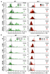
Figure 5. Long Telomeres Mark a Subset of Proliferating Cardiac Cells, Including Proliferating Cardiomyocytes
(A and B) Telomere fluorescence frequency histograms of PCNA and PCNA+ WT and tert−/− cells in uninjured hearts and hearts at 3, 7, 14, 30, and 60 days after cryoinjury. Histograms show frequencies for (A) total cardiac cells and (B) cardiomyocytes. Telomere fluorescence averages are indicated in a.u. Note that in both cases the subpopulation of proliferating cells with long telomeres is lacking in tert−/− hearts. CM, cardiomyocytes. See also Figure S6.
Figure 6
Figure 6. DNA Damage Increases Strongly after Ventricular Cryoinjury in the Absence of Telomerase
(A) Representative western blot of γ-H2AX expression in WT and tert−/− hearts without injury and in hearts at 3 dpi. (B) Quantification of western blot signal intensities (n = 9 hearts/condition). Data are means ± SEM. *p < 0.05 (Mann-Whitney test). (C) Representative staining of γ-H2AX foci (green) in cardiac cells in uninjured and 3 dpi WT and tert−/− hearts. Cardiomyocytes are immunostained with anti-MHC (red), and nuclei are counterstained with DAPI (blue). Examples of γ-H2AX+ cardiomyocytes are outlined with green circles. Boxed areas are shown at a higher magnification. Scale bars, 20 μm. (D) Distribution of γ-H2AX-positive cardiomyocytes (green circles) in uninjured and 3 dpi WT and tert−/− hearts. The nuclear area is shown in magenta. The ventricle and injured area are outlined by dotted lines. Scale bars, 100 μm. (E and F) Percentages of (E) γ-H2AX-positive cardiac cells and (F) γ-H2AX-positive cardiomyocytes in uninjured and 3 dpi WT and tert−/− hearts. Data are means ± SEM of cells counted on a minimum of 3 sections/heart in four hearts. *p < 0.05, **p < 0.01, ***p < 0.001 (Student’s t test). See also Figure S7.
Figure 7
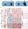
Figure 7. tert−/− Hearts Acquire a Senescence Phenotype after Ventricular Cryoinjury
(A) Whole mounts of uninjured and 3 dpi WT and tert−/− hearts stained for SA β-galactosidase. Scale bars, 100 μm. (B) Cryosections of uninjured, 3 dpi, and 7 dpi WT and tert−/− hearts stained for SA β-galactosidase. Magnified views are shown of the injured area, injury border (B), and remote zone (R). Scale bars, 100 μm (whole heart) and 50 μm (magnified views).

References

    1. Anchelin M, Murcia L, Alcaraz-Pérez F, García-Navarro EM, Cayuela ML. Behaviour of telomere and telomerase during aging and regeneration in zebrafish. PloS ONE. 2011;6:e16955. - PMC - PubMed
    1. Anchelin M, Alcaraz-Pérez F, Martínez CM, Bernabé-García M, Mulero V, Cayuela ML. Premature aging in telomerase-deficient zebra-fish. Dis. Model Mech. 2013;6:1101–1112. - PMC - PubMed
    1. Aurora AB, Porrello ER, Tan W, Mahmoud AI, Hill JA, Bassel-Duby R, Sadek HA, Olson EN. Macrophages are required for neonatal heart regeneration. J. Clin. Invest. 2014;124:1382–1392. - PMC - PubMed
    1. Behrens A, van Deursen JM, Rudolph KL, Schumacher B. Impact of genomic damage and ageing on stem cell function. Nat. Cell Biol. 2014;16:201–207. - PMC - PubMed
    1. Benjamini Y, Hochberg Y. Controlling the false discovery rate: a practical and powerful approach to multiple testing. J. R. Stat. Soc. Series B Stat. Methodol. 1995;57:289–300.

Publication types

Associated data