. 2007 Apr 16;204(4):759-69.
doi: 10.1084/jem.20061923. Epub 2007 Apr 9.

Enhancement and suppression of signaling by the conserved tail of IgG memory-type B cell antigen receptors

Affiliations

Enhancement and suppression of signaling by the conserved tail of IgG memory-type B cell antigen receptors

Keisuke Horikawa et al. J Exp Med. .

Abstract

Immunological memory is characterized by heightened immunoglobulin (Ig) G antibody production caused in part by enhanced plasma cell formation conferred by conserved transmembrane and cytoplasmic segments in isotype-switched IgG B cell receptors. We tested the hypothesis that the IgG tail enhances intracellular B cell antigen receptor (BCR) signaling responses to antigen by analyzing B cells from Ig transgenic mice with IgM receptors or chimeric IgMG receptors containing the IgG tail segment. The IgG tail segment enhanced intracellular calcium responses but not tyrosine or extracellular signal-related kinase (ERK) phosphorylation. Biochemical analysis and crosses to CD22-deficient mice established that IgG tail enhancement of calcium and antibody responses, as well as marginal zone B cell formation, was not due to diminished CD22 phosphorylation or inhibitory function. Microarray profiling showed no evidence for enhanced signaling by the IgG tail for calcium/calcineurin, ERK, or nuclear factor kappaB response genes and little evidence for any enhanced gene induction. Instead, almost half of the antigen-induced gene response in IgM B cells was diminished 50-90% by the IgG tail segment. These findings suggest a novel "less-is-more" hypothesis to explain how switching to IgG enhances B cell memory responses, whereby decreased BCR signaling to genes that oppose marginal zone and plasma cell differentiation enhances the formation of these key cell types.


Figures

Figure 1.
Figure 1.
IgG membrane tail BCR induces exaggerated Ca2+ influx. RBC-depleted splenocytes from IgMG (continuous line) and IgM (dotted line) mice were labeled with 1 μM Indo-1 for 30 min at 37°C. The cells were counterstained with antibodies against B220 and CD21 for the final 10 min of Indo-1 staining. The stained cells were first acquired for 30 s on an LSR flow cytometer and then stimulated with anti-IgM F(ab′)2 antibody (A, 50 μg/ml; B, 5 μg/ml; and C, 0.5 μg/ml) or HEL (D, 5 μg/ml; and E, 0.5 μg/ml; both represent saturating concentrations), and the increase in intracellular calcium was revealed by the change in the mean Indo-1 violet/blue ratio of the B220+CD21medium follicular B cell population. Data are representative of three experiments.
Figure 2.
Figure 2.
IgG membrane tail does not alter tyrosine phosphorylation of CD22 or ERK phosphorylation. Splenocytes from IgM and IgMG transgenic mice were stimulated with 50 μg/ml anti-IgM F(ab′)2. Total cellular proteins (A) or immunoprecipitated CD22 (B) were fractionated by SDS-PAGE and Western blotted with antiphosphotyrosine antibody (A and B, top) or anti-CD22 antibody (B, bottom). The ratios of phosphorylated CD22 to total CD22 are indicated. (C and D) Mean fluorescence intensity (MFI) of permeabilized B220+CD21mediumCD23+ follicular B cells stained by flow cytometry for phosphorylated ERK either (C) at the indicated times after stimulation with 50 μg/ml anti-IgM F(ab′)2 or (D) in unstimulated (−) versus stimulated (+) cells after 2 min in the presence of MEK inhibitors PD98059, U0126, or DMSO as diluent controls. Data are representative of two experiments.
Figure 3.
Figure 3.
The IgG tail does not confer an enhanced gene expression response but blunts a subset of the IgM response. IgM and IgMG follicular B cells were stimulated (S) with 5 mg HEL for 1 h in vivo or were unstimulated (U) and then flow sorted based on CD21 and CD23 staining. Purified RNA from multiple experiments was combined into two independent pools for each stimulation condition and analyzed on Affymetrix U74A microarrays. Increased and decreased genes were defined as those with more than twofold induction or suppression, respectively, and present in both stimulated samples or in both unstimulated samples, respectively. (A and B) Schematic comparing the total number and overlap of genes with increased (A) or decreased (B) expression upon antigen stimulation. The sizes of squares indicate the number of genes. (C) Expression of genes previously defined as induced by HEL stimulation of naive IgM/IgD B cells (references 22, 23). The left end of the line represents the signal of gene expression in unstimulated cells; the right end represents the same in cells stimulated with HEL for 1 h. Each line represents one experiment. Blue line (crosses), IgM B cells; red line (circles), IgMG B cells. (D and E) Hierarchical clustering on all induced (D) or suppressed (E) genes. Red denotes the increased expression relative to the average of all samples, whereas green denotes decreased expression relative to the average. Black denotes values near the average of all samples. Note that approximately half of the response genes in stimulated IgM B cells are induced to lower levels in IgMG B cells.
Figure 4.
Figure 4.
IgG membrane tail evokes an augmented Ca2+ response independent of CD22. RBC-depleted splenocytes from mice of the indicated genotypes were labeled with 1 μM Indo-1 for 30 min at 37°C. (A and B) The cells were counterstained with antibodies against B220 and CD21 for the final 10 min of Indo-1 loading. The stained cells were at first acquired for 30 s and then stimulated with the indicated concentrations of anti-IgM F(ab′)2 antibody (A, 5 μg/ml; and B, 0.5 μg/ml). Lines show the mean Indo-1 ratio in B220+CD21medium cells. Data from one out of three independent experiments are shown. (C and D) Indo-1–loaded cells stained with antibody against B220 were simulated with the indicated concentrations of HEL conjugated to PE (HEL-PE; C, 5 μg/ml; and D, 0.5 μg/ml), and the Indo-1 ratio was measured on gated HEL+ B220+ cells.
Figure 5.
Figure 5.
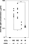
IgG membrane tail increases antibody production independent of CD22. 5 × 10⁵ HEL-binding splenic B cells from IgMG or IgM transgenic donors of the indicated CD22 genotypes were adoptively transferred into nonirradiated C57BL/6 mice, and the recipient mice were immunized with HEL in CFA. The concentration of anti-HEL IgMa antibody 10 d after immunization was measured in the serum of individual recipient mice (circles) by ELISA. Data are representative of three separate experiments. Significant differences were determined by the Mann-Whitney test. *, P < 0.05; **, P < 0.01.
Figure 6.
Figure 6.
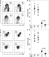
CD22 deficiency suppresses IgM but not IgMG marginal zone B cell differentiation. Splenocytes were stained with HEL and combinations of antibodies against CD21/CD23/HyHEL9/B220 (A and B) or CD21/CD1d/HyHEL9/B220 (C and D). (A and C) The displayed profiles are gated on B220+ HEL-binding cells, and the percentage of B220+ HEL-binding cells in each window is shown. (B and D) Percentages of HEL-binding B cells in the marginal zone subset in individual mice (circles). Significant differences, as determined by the Student's t test, are indicated.

References

    1. Ahmed, R., and D. Gray. 1996. Immunological memory and protective immunity: understanding their relation. Science. 272:54–60. - PubMed
    1. Gray, D. 1993. Immunological memory. Annu. Rev. Immunol. 11:49–77. - PubMed
    1. Reth, M. 1992. Antigen receptors on B lymphocytes. Annu. Rev. Immunol. 10:97–121. - PubMed
    1. Achatz, G., L. Nitschke, and M.C. Lamers. 1997. Effect of transmembrane and cytoplasmic domains of IgE on the IgE response. Science. 276:409–411. - PubMed
    1. Kaisho, T., F. Schwenk, and K. Rajewsky. 1997. The roles of gamma 1 heavy chain membrane expression and cytoplasmic tail in IgG1 responses. Science. 276:412–415. - PubMed

Publication types

MeSH terms