. 2007 May 22;104(21):8953-8.
doi: 10.1073/pnas.0702975104. Epub 2007 May 16.

NRAS mutation causes a human autoimmune lymphoproliferative syndrome

Affiliations

NRAS mutation causes a human autoimmune lymphoproliferative syndrome

João B Oliveira et al. Proc Natl Acad Sci U S A. .

Abstract

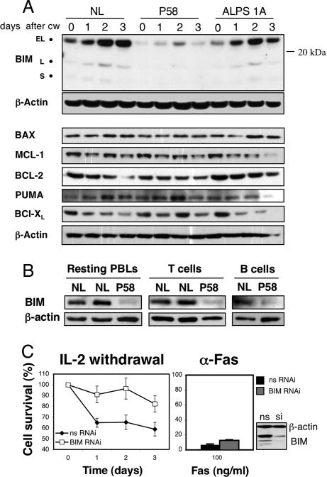
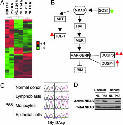
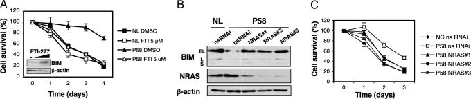
The p21 RAS subfamily of small GTPases, including KRAS, HRAS, and NRAS, regulates cell proliferation, cytoskeletal organization, and other signaling networks, and is the most frequent target of activating mutations in cancer. Activating germline mutations of KRAS and HRAS cause severe developmental abnormalities leading to Noonan, cardio-facial-cutaneous, and Costello syndrome, but activating germline mutations of NRAS have not been reported. Autoimmune lymphoproliferative syndrome (ALPS) is the most common genetic disease of lymphocyte apoptosis and causes autoimmunity as well as excessive lymphocyte accumulation, particularly of CD4(-), CD8(-) alphabeta T cells. Mutations in ALPS typically affect CD95 (Fas/APO-1)-mediated apoptosis, one of the extrinsic death pathways involving TNF receptor superfamily proteins, but certain ALPS individuals have no such mutations. We show here that the salient features of ALPS as well as a predisposition to hematological malignancies can be caused by a heterozygous germline Gly13Asp activating mutation of the NRAS oncogene that does not impair CD95-mediated apoptosis. The increase in active, GTP-bound NRAS augments RAF/MEK/ERK signaling, which markedly decreases the proapoptotic protein BIM and attenuates intrinsic, nonreceptor-mediated mitochondrial apoptosis. Thus, germline activating mutations in NRAS differ from other p21 Ras oncoproteins by causing selective immune abnormalities without general developmental defects. Our observations on the effects of NRAS activation indicate that RAS-inactivating drugs, such as farnesyltransferase inhibitors should be examined in human autoimmune and lymphocyte homeostasis disorders.


Conflict of interest statement

The authors declare no conflict of interest.

Figures

Fig. 1.
Fig. 1.
Defective cytokine withdrawal-induced apoptosis in P58 lymphocytes. (A–D) Activated peripheral blood mononuclear cells (PBLs) from normal volunteers (NL A and B), from a patient with an inactivating Fas mutation (ALPS 1A), and from P58 were cultured in media without IL-2 for the indicated periods of time (A); or treated for 18 h with anti-Fas (Apo1.3) antibody (B), staurosporine (C), or γ-irradiation (D) at the indicated doses. (E) (a–d) Merged views of active BAX (green) and Hoechst nuclear staining (blue) of cells from P58 and a normal control (NL) at 0 h (a and b) and 72 h after cytokine withdrawal (c and d). (e–h) Merged views of staining with the mitochondrial marker Hsp60 (green), cytochrome c (red), and Hoechst staining (blue) at 0 h (e and f) or 72 h after IL-2 withdrawal (g and h). (F) Quantitation using fluorescence microscopy of the relative proportion of cells in the experiment in E showing active BAX expression (activ. BAX), diffuse cytochrome c (diff. cyt. c), or apoptotic (apop.) nuclei. Data shown are the representative of two or three independent experiments. Shown is mean ± SD.
Fig. 2.
Fig. 2.
BIM down-regulation in P58 lymphocytes. (A) Analysis by immunoblotting of BIM and other BCL-2 members expression after IL-2 withdrawal (CW) in PBLs from a NL, P58, and an ALPS 1A patient. The isoforms extra-long (EL), long (L), and short (S) are indicated. β-Actin is a loading control. (B) Resting ex vivo PBLs and purified T and B cells from normals (NL) and P58 were lysed and assessed for BIM expression by immunoblotting. (C) Activated human lymphocytes were transfected with either nonsilencing RNAi (nsRNAi) or with a small interfering oligonucleotide directed at BIM (BIM RNAi) for 3 days, and then deprived of IL-2 or treated with anti-Fas antibody. Silencing efficiency was assessed by immunoblotting (Lower Right) for the nonspecific (ns) and the silencing (si) transfections. Data shown are the representative of three independent experiments. Shown is mean ± SD.
Fig. 3.
Fig. 3.
Identification of a de novo gain-of-function NRAS mutation in P58. (A) Heat map and relational dendrogram results of a microarray study demonstrating the 205 probe sets differentially expressed by cells from P58 compared with cells from two normal subjects, NL1 and NL2. Activated lymphocytes were lysed at 0 and 24 h after IL-2 withdrawal, and mRNA expression was analyzed by using oligonucleotide microarrays (Affymetrix U133Plus2.0). (B) Simplified diagram demonstrating differential expression in P58 cells of genes in the NRAS/RAF/ERK pathway. Red and green arrows indicate the genes up- or down-regulated in P58, respectively, compared with controls. (C) Sequencing of NRAS by using genomic DNA from P58 lymphoblasts, monocytes, and buccal epithelial cells, all demonstrating a heterozygous G-to-A substitution, which changes codon 13 from glycine to aspartic acid. (D) Active GTP-bound NRAS was immunoprecipitated before and after serum withdrawal in a NL and P58, by using a Raf-1 (RBD)-GST fusion protein as bait. The total quantity of NRAS in cell lysates before immunoprecipitation is also shown.
Fig. 4.
Fig. 4.
BIM down-regulation by NRAS through ERK. (A and B) Expression constructs for hemagglutinin (HA)-tagged wild type (WT) or active mutant (G13D) NRAS were transfected into the human lymphoid cell line H-9 (A) or primary human activated lymphocytes (B), followed 48 h later by quantification of BIM expression by immunoblotting. The numbers beneath each lane are the relative intensities of the bands in arbitrary units compared to the corresponding control lane. (C) Activated lymphocytes from two NL and from P58 were deprived of IL-2 and treated with either the DMSO vehicle or MEK1 inhibitors PD98059 (PD) (20 μM) or U0126 (10 μM) for 18 h, after which expression of BIM and BAX were analyzed by immunoblotting. (D) Lymphocytes from P58 and a normal control (NL) were deprived of IL-2 and treated daily with DMSO or PD98059 (20 μM). Apoptosis was measured daily by flow cytometry. Data shown are representative of three or more independent experiments. Shown is mean ± SD. EV, empty vector.
Fig. 5.
Fig. 5.
Correction of the apoptotic defect in P58 by inhibition of farnesylation and RNA silencing. (A) Activated lymphocytes from P58 and a normal control (NL) were deprived of IL-2 and treated daily with DMSO or FTI-277 (5 μM). (Inset) Resting lymphocytes from P58 were treated with DMSO (−) or 2, 5, or 10 μM FTI-277, and BIM expression was analyzed by immunoblotting. (B) Activated lymphocytes from a NL and P58 were transfected with nonsilencing (nsRNAi) or three different NRAS-targeted siRNA oligonucleotides (NRAS#1/2/3), and BIM expression was measured 3 days later by immunoblotting. (C) Activated lymphocytes from a NL and P58 were transfected as described in B and subjected to IL-2 withdrawal, and apoptosis was measured daily for the indicated period of time. Data shown are representative of three or more independent experiments. Shown is mean ± SD.

References

    1. Malumbres M, Barbacid M. Nat Rev Cancer. 2003;3:459–465. - PubMed
    1. Barbacid M. Annu Rev Biochem. 1987;56:779–827. - PubMed
    1. Aoki Y, Niihori T, Kawame H, Kurosawa K, Ohashi H, Tanaka Y, Filocamo M, Kato K, Suzuki Y, Kure S, et al. Nat Genet. 2005;37:1038–1040. - PubMed
    1. Rodriguez-Viciana P, Tetsu O, Tidyman WE, Step AL, Conger BA, Cruz MS, McCormick F, Paven KA. Science. 2006;3:1287–1290. - PubMed
    1. Schubbert S, Zenker M, Rowe SL, Boll S, Klein C, Bollag G, van der Burgt I, Musante L, Kalscheuer V, Wehner LE, et al. Nat Genet. 2006;38:331–336. - PubMed

Publication types

MeSH terms

Associated data