. 2015 Nov 3;112(44):13717-22.
doi: 10.1073/pnas.1510595112. Epub 2015 Oct 19.

Transgenic expression of the proneural transcription factor Ascl1 in Müller glia stimulates retinal regeneration in young mice

Affiliations

Transgenic expression of the proneural transcription factor Ascl1 in Müller glia stimulates retinal regeneration in young mice

Yumi Ueki et al. Proc Natl Acad Sci U S A. .

Abstract

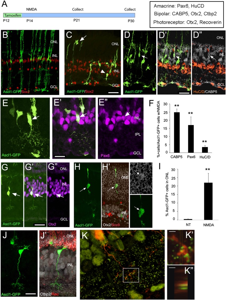
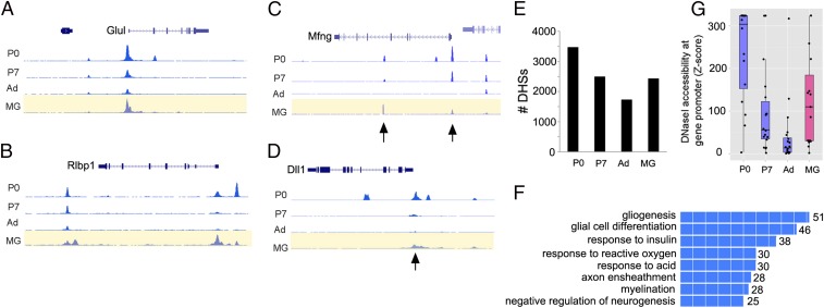
Müller glial cells are the source of retinal regeneration in fish and birds; although this process is efficient in fish, it is less so in birds and very limited in mammals. It has been proposed that factors necessary for providing neurogenic competence to Müller glia in fish and birds after retinal injury are not expressed in mammals. One such factor, the proneural transcription factor Ascl1, is necessary for retinal regeneration in fish but is not expressed after retinal damage in mice. We previously reported that forced expression of Ascl1 in vitro reprograms Müller glia to a neurogenic state. We now test whether forced expression of Ascl1 in mouse Müller glia in vivo stimulates their capacity for retinal regeneration. We find that transgenic expression of Ascl1 in adult Müller glia in undamaged retina does not overtly affect their phenotype; however, when the retina is damaged, the Ascl1-expressing glia initiate a response that resembles the early stages of retinal regeneration in zebrafish. The reaction to injury is even more pronounced in Müller glia in young mice, where the Ascl1-expressing Müller glia give rise to amacrine and bipolar cells and photoreceptors. DNaseI-seq analysis of the retina and Müller glia shows progressive reduction in accessibility of progenitor gene cis-regulatory regions consistent with the reduction in their reprogramming. These results show that at least one of the differences between mammal and fish Müller glia that bears on their difference in regenerative potential is the proneural transcription factor Ascl1.

Keywords: eye; glia; neurogenesis; regeneration; reprogramming.


Conflict of interest statement

The authors declare no conflict of interest.

Figures

Fig. 1.
Fig. 1.
Transgenic Ascl1 overexpression reprograms Müller glia in vitro. (A and B) Diagrams showing the strategy for transgenic Ascl1 overexpression using either AlphaPax6-cre (for retinal-specific expression), rtTAfl-stop, and tetO-Ascl1-IRES-mCherry (hAscl1-mCherry); or Glast-creER (for glial-specific expression), tTAfl-stop, and tetO-Ascl1-IRES-GFP (Ascl1-GFP). (C) Bar plot of RT-PCR–cycle difference from control showing means and SEM for Ascl1, progenitor genes (black), or glial genes (blue) in P12 retinal explants. n = 4; *P < 0.01, **P < 0.005, ***P < 0.0001. (D) Dissociated culture of P12 control and Ascl1-GFP Müller glia, maintained in vitro for 11 d; labeled for antibodies as shown. (Scale bar, 50 μm.) (E) Sections from control mice (CC-GFP = Cag-Cat-eGFP) that received intraperitoneal injections of tamoxifen as adults, colabeled for Sox9 (red). (F) Section through adult mouse retina after 3 wk of Ascl1-GFP expression in the Müller glia, colabeled for Sox9 (white) and Otx2 (red). (Scale bars for E and F, 20 μm.)
Fig. 2.
Fig. 2.
Transgenic Ascl1 overexpression reprograms Müller glia after injury. (A) Diagram showing the experimental design. (B) Retinal section of adult Ascl1- GFP mouse after tamoxifen injections, followed by a single intraocular injection of NMDA, and collected 1 wk later. Anti-GFAP (white) shows Müller glial reactivity after the NMDA (B′, B′′, and D). Müller glia in the Ascl1-GFP retina migrate to the ONL (arrows). (Scale bar, 20 μm.) Red shows anti-Sox9. (C and D) Retinal sections of CC-GFP mice that received no treatment (NT, C) or NMDA (D). The Müller glia retain Sox9 (red) expression in controls and do not migrate into the ONL. (E) Ascl1-GFP–expressing Müller glia express Otx2 (red, arrowheads) and down-regulate Sox9 (white, arrows). (Scale bar, 20 μm.) (F) Graph showing the percentages of Sox9+ and Otx2+ Müller glia in CC-GFP and Ascl1-GFP retinas and in Ascl1-GFP+ retinas following NMDA or light damage. The increase in Otx2 expression after damage in the Ascl1-GFP retinas was significant by a Student’s t test at P = 0.027 (n = 3 for each group). (G) Ascl1-GFP+ light-damaged retina showing Sox9+ (red) Müller glia expressing the mitotic cell-cycle marker, Phospho-Histone3 (white) (arrows). (H) Section through the retinas of light-damaged, Otx2+ progeny of Ascl1-GFP+ Müller glia migrate to the ONL. (Boxed region, Right) Ascl1-GFP+ Müller glia express Otx2. (Scale bar, 20 μm.)
Fig. 3.
Fig. 3.
(A) Diagram showing the experimental design for Ascl1 transgenic expression in Müller glia in young mice. (B) Retinal section through P21 Ascl1- GFP mouse that received tamoxifen at P12. All labeled cells are Sox9+ (red) and have Müller glial morphology. (CE, G, H, J, and K) Retinal sections through P21 Ascl1-GFP mouse that received tamoxifen at P12 and a single intraocular injection of NMDA at P14. (C) The Ascl1-GFP+ cells now take on a variety of different morphologies and migrate to the ONL (arrows) and inner part of the INL (arrowhead). (DD′′) Example of a CABP5+ bipolar cell (arrow). (EE′′) Example of Pax6+ amacrine cell. (F) Percentage of Ascl1-GFP+ cells that express the marker shown. Significance compared with undamaged Ascl1-GFP+ retinas, where no cells with these labels were observed (**P < 0.001, n = 5) (G) Example of Otx2+ bipolar cell derived from Müller glia. (H) Example of Otx2+ (white) rod photoreceptor-like cell, negative for Sox9 (red). (Inset) The Otx2 labeling in the GFP+ cell has acquired the rod-like morphology. (I) Graph of the percent of Ascl1-GFP+ cells in the ONL in undamaged Ascl1-GFP+ retinas and after NMDA damage. **P = 0.0017. (n = 5) (J and J′) Example of bipolar cell derived from Ascl1-GFP+ Müller glia, showing CtBP2 labeling (white) and recoverin (red) labeled photoreceptors and termini in the OPL. (K, K′, K′′) Overlap between CtBP2 (green) and GFP (red) in a Müller glial-derived bipolar cell. (Scale bars, 1 μm.)
Fig. 4.
Fig. 4.
DNaseI-seq analysis of Müller glia shows accessible chromatin at progenitor gene loci. (A and B) Examples of Müller glial-expressed genes, glutamate synthetase (Glul), and cellular retinaldehyde-binding protein (Rlbp1) at three stages of retinal development and in P12 Müller glia. (C and D) Examples of progenitor genes, Mfng and Dll1, in P12 Müller glia. Arrows point to peaks present in P0 retina that remain accessible at P7 and in P12 Müller glia. (E) P0 DHSs that overlap with Ascl1-ChIP-seq in neural progenitors that are also present at P7, adult retina and P12 Müller glia. (F) The P0-DHS/Ascl1-ChIP-seq peaks DHSs shared with Müller glia are enriched for GO Biological processes associated with neural developmental genes. (G) The degree of DNase1 hypersensitivity at the promoters of 15 progenitor genes in P0, P7, or adult retina, compared with Müller glia, using z scores (e−x). Box plots with means and data points shown.

References

    1. Goldman D. Müller glial cell reprogramming and retina regeneration. Nat Rev Neurosci. 2014;15(7):431–442. - PMC - PubMed
    1. Lenkowski JR, Raymond PA. Müller glia: Stem cells for generation and regeneration of retinal neurons in teleost fish. Prog Retin Eye Res. 2014;40:94–123. - PMC - PubMed
    1. Karl MO, Reh TA. Regenerative medicine for retinal diseases: Activating endogenous repair mechanisms. Trends Mol Med. 2010;16(4):193–202. - PMC - PubMed
    1. Karl MO, Reh TA. Studying the generation of regenerated retinal neuron from Müller glia in the mouse eye. Methods Mol Biol. 2012;884:213–227. - PubMed
    1. Fischer AJ, Reh TA. Müller glia are a potential source of neural regeneration in the postnatal chicken retina. Nat Neurosci. 2001;4(3):247–252. - PubMed

Publication types

MeSH terms

Substances

Associated data

LinkOut - more resources