. 2005 Jun 9;435(7043):828-33.
doi: 10.1038/nature03552.

A microRNA polycistron as a potential human oncogene

Affiliations

A microRNA polycistron as a potential human oncogene

Lin He et al. Nature. .

Abstract

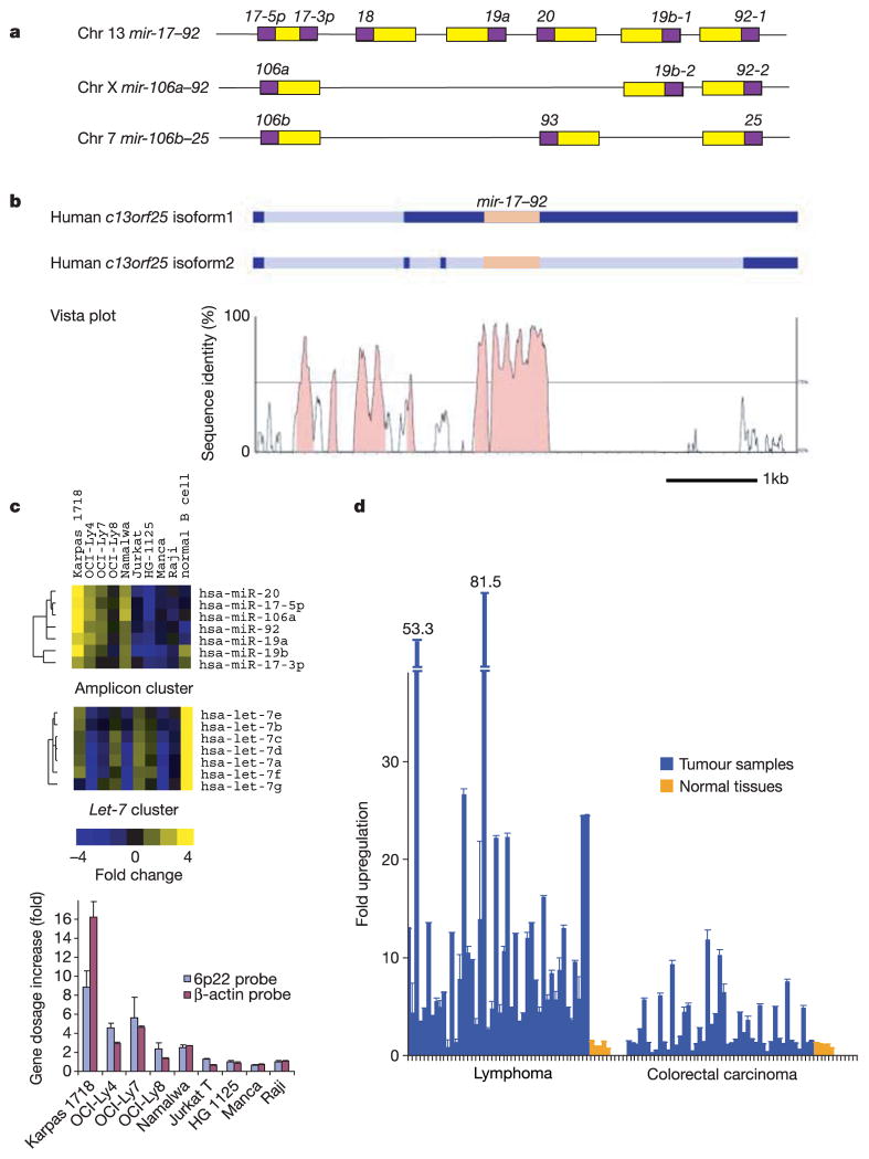
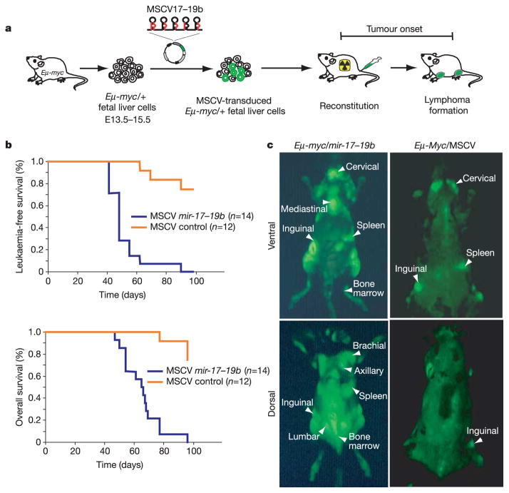
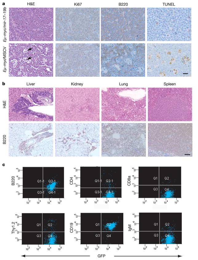
To date, more than 200 microRNAs have been described in humans; however, the precise functions of these regulatory, non-coding RNAs remains largely obscure. One cluster of microRNAs, the mir-17-92 polycistron, is located in a region of DNA that is amplified in human B-cell lymphomas. Here we compared B-cell lymphoma samples and cell lines to normal tissues, and found that the levels of the primary or mature microRNAs derived from the mir-17-92 locus are often substantially increased in these cancers. Enforced expression of the mir-17-92 cluster acted with c-myc expression to accelerate tumour development in a mouse B-cell lymphoma model. Tumours derived from haematopoietic stem cells expressing a subset of the mir-17-92 cluster and c-myc could be distinguished by an absence of apoptosis that was otherwise prevalent in c-myc-induced lymphomas. Together, these studies indicate that non-coding RNAs, specifically microRNAs, can modulate tumour formation, and implicate the mir-17-92 cluster as a potential human oncogene.


Conflict of interest statement

The authors declare no competing financial interests.

Figures

Figure 1
Figure 1. The mir-1792 cluster shows increased expression in B-cell lymphoma samples and cell lines
a, Genomic organization of three polycistronic miRNA clusters is shown. There are five paralogous groups located in three homologous clusters (mir-17–92, mir-106a–92 and mir-106b–25) with a conserved order: miR-17-5p/miR-106a/miR-106b; miR-18; miR-19a/miR-19b-1/miR-19b-2; miR-20/miR-93; and miR-92-1/miR-92-2/miR-25 (yellow boxes, pre-miRNAs; purple boxes, mature miRNAs). b, The level of conservation between human and mouse homologues is represented using an mVista plot. Two alternative isoforms have been detected for the human gene, and these are shown schematically (dark blue, exons; light blue, introns; orange, the mir-1792 cluster). c, MicroRNA expression levels in cell lines carrying the 13q31-q32 amplicon (including Karpas 1718, OCI-Ly4 and OCI-Ly7) were compared to those in leukaemia and lymphoma cell lines lacking this genetic lesion, and to normal B-cells isolated from cortical blood (top panel). We included in this analysis the OCI-Ly8 cell line, which has previously been identified as a cell line carrying the 13q31-32 amplicon, but showed no gene dosage increase at the c13orf25 locus in our study. Normalized one-channel measurements for 191 human miRNAs were hierarchically clustered for all miRNA genes represented on the array. An excerpt of the data is shown, and the full cluster analysis is presented in Supplementary Fig. 1. The expression map node that correlates with the amplification is shown. The let-7 cluster node is also shown for comparison (middle panel). In the cell lines examined, expression levels of the mature microRNAs from the mir-1792 polycistron correlate with the copy number at the mir-1792 locus (bottom panel). d, The level of mir-1792 pri-miRNA was determined by real-time quantitative PCR in 46 lymphomas and 47 colorectal carcinomas, and compared to levels found in corresponding normal tissues from five individuals. In c and d, error bars indicate standard deviation (s.d.).
Figure 2
Figure 2. Overexpression of the mir-1719b cluster accelerates c-myc-induced lymphomagenesis in mice
a, Schematic representation of the adoptive transfer protocol using -myc HSCs. b, Mice reconstituted with HSCs expressing mir-1719b in an MSCV retroviral vector (MSCV mir-1719b) or infected with a control MSCV virus were monitored by blood smear analysis starting 5 weeks after transplantation. The Kaplan-Meier curves represent the percentage of leukaemia-free survival or overall survival. c, External GFP imaging of tumour-bearing mice with -myc/mir-1719b or -myc/MSCV shows the overall distribution of tumour cells. -myc/mir-1719b tumours show a more disseminated phenotype compared with control tumours. These animals are representative of their genotype.
Figure 3
Figure 3. Pathological and immunological analysis of lymphomas produced by cooperation between mir-1719b and c-myc
a, Haemotoxylin and eosin (H&E), Ki67, B220 and TUNEL staining of -myc/mir-1719b lymph node tumours. The ‘starry sky’ morphology is a hallmark of cell clusters undergoing apoptosis (black arrows). Scale bar, 10μm. b, Invasion of visceral organs (liver, spleen, lung and kidney) by -myc/mir-1719b tumour cells, shown by H&E and B220 staining. Invasion was observed both perivascularly and parenchymally in liver. Scale bar, 50μm. c, Immunophenotyping of -myc/mir-1719b lymphomas. Tumour cells stained positively for the B-cell-specific marker B220, but not for the T-cell-specific markers CD4, CD8a and Thy1.2. Tumour cells bore cellular characteristics of pre-B cells, staining positively for CD19 but not for IgM, a marker of mature B-cells.

Comment in

References

    1. Ota A, et al. Identification and characterization of a novel gene, C13orf25, as a target for 13q31-q32 amplification in malignant lymphoma. Cancer Res. 2004;64:3087–3095. - PubMed
    1. Lee Y, et al. The nuclear RNase III Drosha initiates microRNA processing. Nature. 2003;425:415–419. - PubMed
    1. Bernstein E, Caudy AA, Hammond SM, Hannon GJ. Role for a bidentate ribonuclease in the initiation step of RNA interference. Nature. 2001;409:363–366. - PubMed
    1. Bartel DP. MicroRNAs: genomics, biogenesis, mechanism, and function. Cell. 2004;116:281–297. - PubMed
    1. Ambros V. The functions of animal microRNAs. Nature. 2004;431:350–355. - PubMed

Publication types

Associated data