. 2025 Jun 4;15(1):19633.
doi: 10.1038/s41598-025-03589-1.

Analysis of human factor H-related gene and protein expressed in rheumatoid arthritis synovium identifies a novel mechanism promoting dysregulated complement pathway activation

Collaborators, Affiliations

Analysis of human factor H-related gene and protein expressed in rheumatoid arthritis synovium identifies a novel mechanism promoting dysregulated complement pathway activation

Nirmal K Banda et al. Sci Rep. .

Abstract

Factor H (FH) is a negative regulator of the alternative pathway (AP) of complement however, five human factor H-related (FHR) proteins, can also function ex vivo as positive regulators. We compare bulk FH and FHR mRNA expressions in both the human rheumatoid arthritis (RA) synovium and blood cells from the Pathobiology of Early Arthritis Cohort (PEAC) and the Stratification of biological therapies for Rheumatoid Arthritis by Pathobiology (STRAP) Cohort. FH and FHR proteins were detected using multiplexed immunohistochemistry (MIHC) in synovium. In three pathotypes, in the synovium, no differences were found in the expression of FHR mRNA. In the synovium, a significant negative correlation was observed between FH expression and the disease activity score and X-ray joint space narrowing. In RA patients, there was a significant positive correlation between FHR3 mRNA level, anti-cyclic citrullinated peptide (CCP) antibodies and rheumatoid factor (RF). FHR proteins were co-localized in the synovial lining area along with complement C3 while FH was almost undetectable in the synovial lining but abundant in sub-synovial lining areas. We do not know whether FH and FHR proteins are locally generated and deposited in synovium or come from circulation. In sum, due to the absence of FH but the presence of FHRs, the synovial lining might fail to be protected from complement-mediated attack, and FHR3 may play a particularly important pathogenic role.


Conflict of interest statement

Declarations. Competing interests: The authors declare no competing interests. NKB: royalties, and patents for the treatment of inflammatory diseases using anti-C5aR1ab-C5siRNA conjugate. LWM: none; KDD: none; DS, none; RIS: none; RMF: none; JS, none; RL: none; CP: none; MJL: none; VMH: royalties, consulting income, stock, and stock options in complement therapeutics companies. Ethical approval and consent to participate: Written ethical informed consent was obtained from all eRA patients according to the preapproved Colorado Multiple Institutional Review Board protocol (COMIRB # 20-1908 and 15-1389). All methods were carried out in accordance with relevant guidelines and regulations. All experimental protocols related to these studies were approved by the Institutional Review Board committee.

Figures

Fig. 1
Fig. 1
A representative composite image from nine eRA synovial samples generated after MIHC showing the presence of all five human FHR proteins along with complement component C3c and membrane attack complex (a.k.a. C5b-9). A The presence of FHR1 (magenta color), FHR2 (white color), FHR3 (green color), FHR4 (pink color), FHR5 (yellow color), FH (cyan color), C3c (red color) and MAC (C5b-9) (orange color) has been shown in the synovial biopsies. B FHR2 and FHR3 compared with FH dominantly present in the synovial lining of synovial biopsies. A false color-coding key was used for each complement protein shown in this composite image. Adipocytes = black cobble-like stone cells in the synovium. A minimum of three to five composite images were generated using a single synovial biopsy with 2 sections from each patient. eRA synovial biopsies n = 9. Magnification = × 40. Scale bar = 100 μm. *p < 0.05.
Fig. 2
Fig. 2
Showing percentage of FHR, FH, C3c and MAC single and double positive synovial cells in the synovium from eRA patients. MIHC was used to generate composite images followed by quantitation of single and double positive cells showing the presence of FHR1, FHR2, FHR3, FHR4, FHR5, FH, C3c, and MAC. A Presence of single positive synovial cells expressing FHR1, FHR2, FHR3, FHR4, FHR5, FH, C3c, and MAC. B Presence of double positive synovial cells positively stained for proteins FHR1/C3c, FHR2/C3, FHR3/C3, FHR4/C4, FHR5/C3c, FH/C3c and MAC/C3c. Synovial cells expressed a lower level of FHR1, FHR4, FHR5 and MAC. eRA synovial biopsies n = 8. Data are shown as mean ± SEM. *p < 0.05 considered significant.
Fig. 3
Fig. 3
A representative composite image from eight synovial samples showing the presence of macrophages and fibroblasts expressing FHR proteins in eRA patients. A Synovial cells showing the presence of FHR1 (magenta color), FHR2 (white color), FHR3 (green color), FHR4 (pink color), FHR5 (yellow color), FH (cyan color), DAPI (blue), macrophages (red color) and fibroblasts (orange color). B Synovium cells showing the predominance of FHR3 (green color) in the sub synovial lining area. A false color-coding key was used for each complement protein, macrophages and fibroblasts shown in this composite image. Adipocytes = black cobble-like stone cells in synovium. A minimum of three to five composite images were generated using a single synovial biopsy with 2 sections from each patient. Magnification = × 40. eRA synovial biopsies n = 8. Scale bar = 100 μm.
Fig. 4
Fig. 4
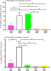
Bar graphs show the presence of macrophages and fibroblasts positive for individual FHR proteins in the synovium from eRA patients. MIHC composite images were analyzed for the presence of percentages of synovial macrophages (CD68 positive) and fibroblasts (FAP) positive for FHR1, FHR2, FHR3, FHR4 and FHR5 proteins. A Percentage of macrophages positive for FHR1, FHR2, FHR3, FHR4 and FHR5 proteins. B Percentage of fibroblasts positive for FHR1, FHR2, FHR3, FHR4 and FHR5 proteins. Both macrophages and fibroblasts have a low level of FHR1, FHR4 and FHR5 proteins compared with FHR2 and FHR3 proteins. eRA synovial biopsies n = 8. Data are shown as mean ± SEM. *p < 0.05 considered significant.
Fig. 5
Fig. 5
Representative snapshots of MIHC composite images showing the regional and differential distribution of FHR proteins in the synovial biopsies from eRA patients. A Composite image showing coexistence of FHR2/FH in the synovial lining but coexistent of FHR5/FH in the sub synovial lining area. B FH is predominately present in the sub synovial lining area, not in the synovial lining area. C FHR1 and FHR4 proteins are positively stained in cells in the synovial lining area. FHR3 is also present in the sub-synovial lining areas. D C3c is predominately and constantly present in the synovial lining cells. The coexistence of FHR1, FHR2, and FHR4 was also noticed in the sub-synovial lining areas. A false color-coding key represents FHR1 = magenta, FHR2 = white, FHR3 = green, FHR4 = pink, FHR5 = yellow, C3c = red, FH = cyan and DAPI = blue. SL = synovial lining. SSL = sub synovial lining Magnification = × 40. eRA synovial biopsies n = 4. Scale bar = 100 μm.
Fig. 6
Fig. 6
Correlations between FHR3, RF, and anti-CCP in the synovium from RA patients from STRAP clinical trial. A Positive and significant correlation between FHR3 gene expression and RF autoantibodies. B Positive and significant correlation between FHR3 gene expression and anti-CCP autoantibodies. RF rheumatoid factor antibody. CCP anti-cyclic citrullinated peptide antibody. STRAP n = 208. Correlation = r and *p < 0.05 are considered significant.
Fig. 7
Fig. 7
Correlations between FHR3, X-ray erosion, and X-ray joint space narrowing from the STRAP clinical trial. A No correlation between FHR3 mRNA expression and X-ray erosion in RA patients. B No correlation between FHR3 mRNA expression and X-ray joint space narrowing in RA patients. C No correlation between FH mRNA expression and X-ray erosion in RA patients. D A negative and significant correlation between FH mRNA expression and X-ray joint space narrowing in RA patients. X-ray erosion = radiographically (X-ray) demonstrable bone erosion in RA. X-ray joint space narrowing = radiographs (X-ray) used to manually measure and quantify joint space to determine the severity of RA. STRAP n = 208. Correlation = r and *p < 0.05 are considered significant.
Fig. 8
Fig. 8
A putative model showing the theoretical inhibition and activation of complement system by FH and FHR proteins respectively on the surface of synovial lining in eRA. A No complement activation in the presence of FH binding to the synovial lining surface bound C3b and sialic acid. B Complement activation in the presence of FHR proteins and absence of FH binding to various complement fragments such as C3b, iC3b, C3dg, and C3d on the surface of the synovial lining. C Complement activation in the presence and binding of various FHR proteins to the synovial lining. All complement fragments, FH, FHR proteins and sialic acid have been color coded. SL = synovial lining (red color arch outer area). Sub synovial lining area FH is present.

References

    1. Cross, M. et al. The global burden of rheumatoid arthritis: estimates from the global burden of disease 2010 study. Ann. Rheumatol. Dis.73, 1316–1322 (2014). - PubMed
    1. Smolen, J. S. et al. Rheumatoid arthritis. Nat. Rev. Dis. Primers. 4, 18001 (2018). - PubMed
    1. Fallon, E. A. et al. Prevalence of diagnosed Arthritis - United States, 2019–2021. MMWR Morb Mortal. Wkly. Rep.72, 1101–1107 (2023). - PMC - PubMed
    1. Singh, D. K. et al. Use of rheumatologic testing in patients who eventually receive a diagnosis of rheumatoid arthritis. South Med. J.112, 535–538 (2019). - PMC - PubMed
    1. Prevoo, M. L., van ‘t Hof, M. A., Kuper, H. H., van Leeuwen, M. A. & van de Putte, L. B. and P. L. van Riel. Modified disease activity scores that include twenty-eight-joint counts. Development and validation in a prospective longitudinal study of patients with rheumatoid arthritis. Arthritis Rheumatol 38: 44–48. (1995). - PubMed