. 2022 Jun 25;8(1):83.
doi: 10.1186/s40798-022-00469-0.

Resuming Training in High-Level Athletes After Mild COVID-19 Infection: A Multicenter Prospective Study (ASCCOVID-19)

Collaborators, Affiliations

Resuming Training in High-Level Athletes After Mild COVID-19 Infection: A Multicenter Prospective Study (ASCCOVID-19)

Laurent Chevalier et al. Sports Med Open. .

Abstract

Background: There is a paucity of data on cardiovascular sequelae of asymptomatic/mildly symptomatic SARS-Cov-2 infections (COVID).

Objectives: The aim of this prospective study was to characterize the cardiovascular sequelae of asymptomatic/mildly symptomatic COVID-19 among high/elite-level athletes.

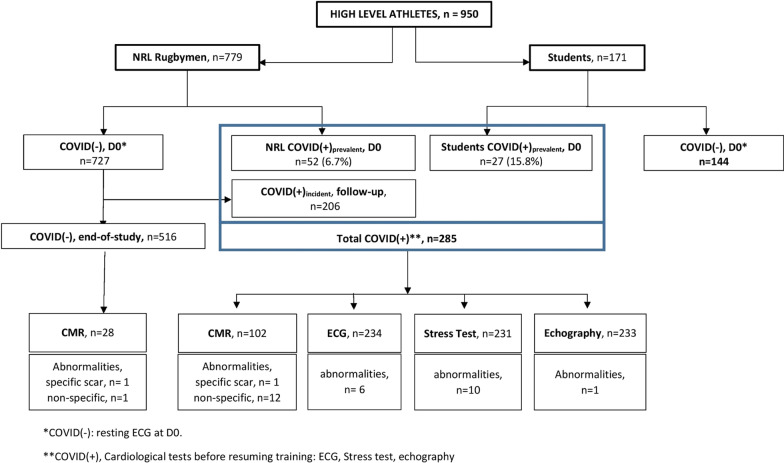
Methods: 950 athletes (779 professional French National Rugby League (F-NRL) players; 171 student athletes) were included. SARS-Cov-2 testing was performed at inclusion, and F-NRL athletes were intensely followed-up for incident COVID-19. Athletes underwent ECG and biomarker profiling (D-Dimer, troponin, C-reactive protein). COVID(+) athletes underwent additional exercise testing, echocardiography and cardiac magnetic resonance imaging (CMR).

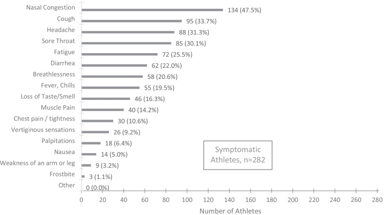
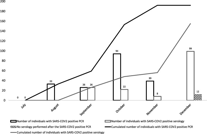
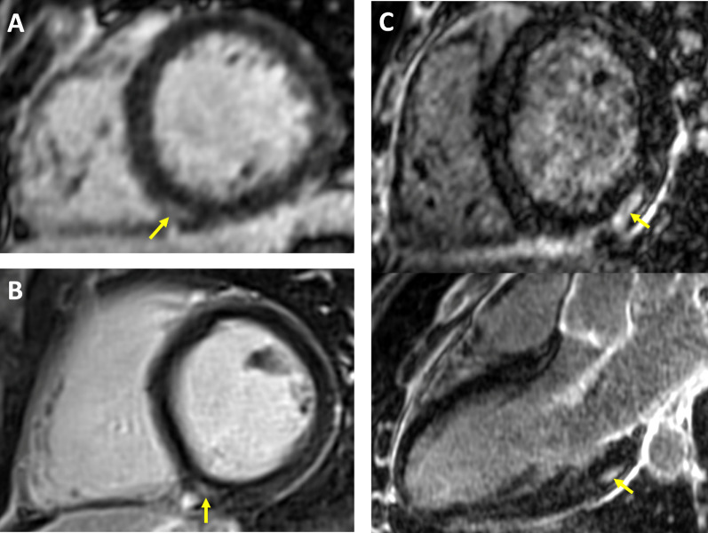
Results: 285/950 athletes (30.0%) had mild/asymptomatic COVID-19 [79 (8.3%) at inclusion (COVID(+)prevalent); 206 (28.3%) during follow-up (COVID(+)incident)]. 2.6% COVID(+) athletes had abnormal ECGs, while 0.4% had an abnormal echocardiogram. During stress testing (following 7-day rest), COVID(+) athletes had a functional capacity of 12.8 ± 2.7 METS with only stress-induced premature ventricular ectopy in 10 (4.3%). Prevalence of CMR scar was comparable between COVID(+) athletes and controls [COVID(+) vs. COVID(-); 1/102 (1.0%) vs 1/28 (3.6%)]. During 289 ± 56 days follow-up, one athlete had ventricular tachycardia, with no obvious link with a SARS-CoV-2 infection. The proportion with troponin I and CRP values above the upper-limit threshold was comparable between pre- and post-infection (5.9% vs 5.9%, and 5.6% vs 8.7%, respectively). The proportion with D-Dimer values above the upper-limit threshold increased when comparing pre- and post-infection (7.9% vs 17.3%, P = 0.01).

Conclusion: The absence of cardiac sequelae in pauci/asymptomatic COVID(+) athletes is reassuring and argues against the need for systematic cardiac assessment prior to resumption of training (clinicaltrials.gov; NCT04936503).

Keywords: Cardiac MRI; ECG; Echocardiography; SARS-CoV-2; Sport; Stress test.


Conflict of interest statement

Abbott has kindly provided the anti-SARS-CoV-2 serology tests (IgG and IgM) to the biology department of the Bordeaux University Hospital (I. Pellegrin, S. Picard, P. Trimoulet) for centralized serologies. The other authors declare that they have no competing interests.

Figures

Fig. 1
Fig. 1
Reported symptoms evocative of SARS-CoV-2 infection in the study population (regardless of SARS-COV-2 test results). Athletes presenting only clinical signs of SARS-COV-2 infection (anosmia or ageusia) with negative serology and PCR were excluded from the analysis as we could not establish the diagnosis with a sufficient level of certainty
Fig. 2
Fig. 2
Occurrence of positive SARS-CoV-2 diagnosis during F-NRL season (RT-PCR and serologies). The F-NRL protocol involved twice-weekly SARS-CoV-2 RT-PCR testing (D614G variant). COVID(+) diagnosis was based on positive serology or a positive SARS-CoV-2 RT-PCR. 192/206 COVID(+)incident cases were diagnosed by positive RT-PCR and 14 by positive serology (without previous positive RT-PCR). SARS-CoV-2 seroconversion (IgM and/or IgG) occurred in 155 (80.7%) of the 192 players diagnosed by RT-PCR, while 25 (13%) did not show detectable anti-SARS-CoV-2 antibodies during the follow-up period
Fig. 3
Fig. 3
SARS-CoV-2 diagnosis and cardiovascular assessment results1 among athletes included in the ASCCOVID study
Fig. 4
Fig. 4
Late gadolinium enhancement CMR findings in athletes following mildly symptomatic COVID-19 infection. Yellow arrows indicate areas of LGE. A-B: small focal intramural LGE in the vicinity of the posterior RV insertion point. This pattern observed in equal proportions of COVID(+) and COVID(−) athletes, and considered as non-specific because suggestive of physiological remodeling. C: Intramural and sub-epicardial LGE lesion on infero-latero-basal LV segment, shown in short axis and 3-chamber views (top and bottom panel, respectively). The patient had experienced a mild symptomatic SARS-CoV-2 infection 65 days prior to the CMR study, with no clinical signs of myocarditis. Echocardiography, ECG and troponin tests were negative at inclusion. This CMR finding being suggestive of a post-inflammatory origin, scarring secondary to COVID-related myocarditis could not be ruled out

References

    1. Guo T, Fan Y, Chen M, et al. Cardiovascular implications of fatal outcomes of patients with coronavirus disease 2019 (COVID-19) JAMA Cardiol. 2020;5(7):811–818. doi: 10.1001/jamacardio.2020.1017. - DOI - PMC - PubMed
    1. Shi S, Qin M, Shen B, et al. Association of cardiac injury with mortality in hospitalized patients with COVID-19 in Wuhan. China JAMA Cardiol. 2020;5(7):802–810. doi: 10.1001/jamacardio.2020.0950. - DOI - PMC - PubMed
    1. Wang D, Hu B, Hu C, et al. Clinical characteristics of 138 hospitalized patients with 2019 novel coronavirus-infected pneumonia in Wuhan. China JAMA. 2020;323(11):1061–1069. doi: 10.1001/jama.2020.1585. - DOI - PMC - PubMed
    1. Liu PP, Blet A, Smyth D, Li H. The science underlying COVID-19: implications for the cardiovascular system. Circulation. 2020;142:68–78. doi: 10.1161/CIRCULATIONAHA.120.047549. - DOI - PubMed
    1. Clerkin KJ, Fried JA, Raikhelkar J, et al. Coronavirus disease 2019 (COVID-19) and cardiovascular disease. Circulation. 2020;141(20):1648–1655. doi: 10.1161/CIRCULATIONAHA.120.046941. - DOI - PubMed

Associated data