

Performance evaluation of automated white matter hyperintensity segmentation algorithms in a multicenter cohort on cognitive impairment and dementia
- PMID: 36713907
- PMCID: PMC9877422
- DOI: 10.3389/fpsyt.2022.1010273
Performance evaluation of automated white matter hyperintensity segmentation algorithms in a multicenter cohort on cognitive impairment and dementia
Abstract
Background: White matter hyperintensities (WMH), a biomarker of small vessel disease, are often found in Alzheimer's disease (AD) and their advanced detection and quantification can be beneficial for research and clinical applications. To investigate WMH in large-scale multicenter studies on cognitive impairment and AD, appropriate automated WMH segmentation algorithms are required. This study aimed to compare the performance of segmentation tools and provide information on their application in multicenter research.
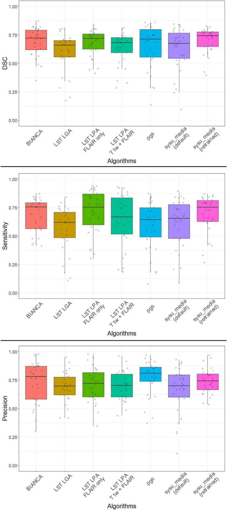
Methods: We used a pseudo-randomly selected dataset (n = 50) from the DZNE-multicenter observational Longitudinal Cognitive Impairment and Dementia Study (DELCODE) that included 3D fluid-attenuated inversion recovery (FLAIR) images from participants across the cognitive continuum. Performances of top-rated algorithms for automated WMH segmentation [Brain Intensity Abnormality Classification Algorithm (BIANCA), lesion segmentation toolbox (LST), lesion growth algorithm (LGA), LST lesion prediction algorithm (LPA), pgs, and sysu_media] were compared to manual reference segmentation (RS).
Results: Across tools, segmentation performance was moderate for global WMH volume and number of detected lesions. After retraining on a DELCODE subset, the deep learning algorithm sysu_media showed the highest performances with an average Dice's coefficient of 0.702 (±0.109 SD) for volume and a mean F1-score of 0.642 (±0.109 SD) for the number of lesions. The intra-class correlation was excellent for all algorithms (>0.9) but BIANCA (0.835). Performance improved with high WMH burden and varied across brain regions.
Conclusion: To conclude, the deep learning algorithm, when retrained, performed well in the multicenter context. Nevertheless, the performance was close to traditional methods. We provide methodological recommendations for future studies using automated WMH segmentation to quantify and assess WMH along the continuum of cognitive impairment and AD dementia.
Keywords: Alzheimer’s disease; FLAIR; aging; deep learning; evaluation; white matter hyperintensities segmentation.
Copyright © 2023 Gaubert, Dell’Orco, Lange, Garnier-Crussard, Zimmermann, Dyrba, Duering, Ziegler, Peters, Preis, Priller, Spruth, Schneider, Fliessbach, Wiltfang, Schott, Maier, Glanz, Buerger, Janowitz, Perneczky, Rauchmann, Teipel, Kilimann, Laske, Munk, Spottke, Roy, Dobisch, Ewers, Dechent, Haynes, Scheffler, Düzel, Jessen and Wirth.
Conflict of interest statement
MDu received fees for consultation and lectures from Roche, Bayer, Hovid Berhad, and Sanofi. OP received fees for consultation and lectures from Biogen, Eisai, Griffols, MSD, Roche, and Schwabe. JP received fees for consultation, lectures, patents from Neurimmune, Axon, Desitin, and Epomedics. JW was an honorary speaker for Actelion, Amgen, Beeijing Yibai Science and Technology Ltd., Janssen Cilag, Med Update GmbH, Pfizer, Roche Pharma, and was a member of the advisory boards of Abbott, Biogen, Boehringer Ingelheim, Lilly, MSD Sharp & Dohme, and Roche Pharma and received fees as a consultant for Immungenetics and Roboscreen. FJ received fees for consultation from Eli Lilly, Novartis, Roche, BioGene, MSD, Piramal, Janssen, and Lundbeck. The remaining authors declare that the research was conducted in the absence of any commercial or financial relationships that could be construed as a potential conflict of interest.
Figures
References
-
- Prins ND, Scheltens P. White matter hyperintensities, cognitive impairment and dementia: an update. Nat Publ Gr. (2015) 11:157–65. - PubMed
LinkOut - more resources
Full Text Sources
Miscellaneous