. 2011 Jul 19;108(29):12113-8.
doi: 10.1073/pnas.1019434108. Epub 2011 Jul 1.

Latrophilin 1 and its endogenous ligand Lasso/teneurin-2 form a high-affinity transsynaptic receptor pair with signaling capabilities

Affiliations

Latrophilin 1 and its endogenous ligand Lasso/teneurin-2 form a high-affinity transsynaptic receptor pair with signaling capabilities

John-Paul Silva et al. Proc Natl Acad Sci U S A. .

Abstract

Latrophilin 1 (LPH1), a neuronal receptor of α-latrotoxin, is implicated in neurotransmitter release and control of presynaptic Ca(2+). As an "adhesion G-protein-coupled receptor," LPH1 can convert cell surface interactions into intracellular signaling. To examine the physiological functions of LPH1, we used LPH1's extracellular domain to purify its endogenous ligand. A single protein of ∼275 kDa was isolated from rat brain and termed Lasso. Peptide sequencing and molecular cloning have shown that Lasso is a splice variant of teneurin-2, a brain-specific orphan cell surface receptor with a function in neuronal pathfinding and synaptogenesis. We show that LPH1 and Lasso interact strongly and specifically. They are always copurified from rat brain extracts. Coculturing cells expressing LPH1 with cells expressing Lasso leads to their mutual attraction and formation of multiple junctions to which both proteins are recruited. Cells expressing LPH1 form chimerical synapses with hippocampal neurons in cocultures; LPH1 and postsynaptic neuronal protein PSD-95 accumulate on opposite sides of these structures. Immunoblotting and immunoelectron microscopy of purified synapses and immunostaining of cultured hippocampal neurons show that LPH1 and Lasso are enriched in synapses; in both systems, LPH1 is presynaptic, whereas Lasso is postsynaptic. A C-terminal fragment of Lasso interacts with LPH1 and induces Ca(2+) signals in presynaptic boutons of hippocampal neurons and in neuroblastoma cells expressing LPH1. Thus, LPH1 and Lasso can form transsynaptic complexes capable of inducing presynaptic Ca(2+) signals, which might affect synaptic functions.


Conflict of interest statement

The authors declare no conflict of interest.

Figures
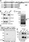
Fig. 1.
Fig. 1.
Isolation of a LPH1 ligand. (A) LPH1-FS and variants of its soluble ectodomains. SP, signal peptide; GBL, galactose-binding lectin; STP, rich in serine, threonine, and proline; GPS, GPCR proteolysis site; V5, V5 epitope. (B) Only LPH-51 binds α-LTX with high affinity. Soluble constructs were expressed in NB2a cells, bound to α-LTX columns, eluted with SDS, and analyzed by immunoblotting with anti-V5 mAb (n = 6). (C) Lasso is only isolated on LPH-51. Detergent extract from rat brain was incubated with V5 Ab/LPH-51 or V5 Ab columns. A control V5 Ab/LPH-51 column was incubated with buffer only. The columns were washed and eluted with alkali. The asterisk denotes the protein (Lasso) isolated on the test column but not on control columns. (D) Lasso is a membrane-bound, brain-specific protein that binds LPH-51 tightly. Three V5 Ab/LPH-51 columns (as in C) were incubated with detergent extracts from rat brain or rat liver or with a buffer extract from rat brain, washed, and eluted with 1 M NaCl and then with alkali. (E) Lasso specifically copurifies with LPH1. Rat brain extract was incubated with an α-LTX column, an anti-LPH1 recombinant Ab (A1) attached to a V5 Ab column, or a BSA column. SDS eluates were analyzed by immunoblotting with the indicated Abs (n = 3).
Fig. 2.
Fig. 2.
Interaction of Lasso with LPH1. (A) Lasso (GenBank accession no. JF784339) and its recombinant constructs. Binding to LPH-51 is summarized on the right. (B) LPH1 binding localizes to the globular domain of Lasso. Soluble constructs (Lasso-B–Lasso-F) were expressed in NB2a cells, incubated with LPH-51, pulled down using FLAG Ab, and analyzed by immunoblotting with anti-V5 Ab. (C) Truncated NTFs of LPH1. Binding to Lasso-D and α-LTX is shown on the right (n = 3). (D) Truncated NTFs do not bind Lasso-D. Soluble LPH-95–LPH-99 were expressed in NB2a cells, incubated with Lasso-D, pulled down using anti-V5 Ab, and analyzed by immunoblotting with FLAG Ab (n = 3). (E) Some NTF constructs bind α-LTX. LPH-95–LPH-99 were attached to microtiter plates via V5 Ab, incubated with α-LTX and detected with anti-α-LTX Ab (mean ± SEM; n = 3). (F) Lasso binds LPH1, but not LPH2 or LPH3. Rat brain lysate was incubated with purified Lasso-D, precipitated with anti-FLAG Ab, and analyzed by immunoblotting with PAL1, PAL2, and PAL3 Abs (36) (n = 2). (G) Characterization of the Lasso–LPH1 interaction. LPH-51 was immobilized on microtiter plates via V5 Ab, incubated with different concentrations of Lasso-D, fixed, and stained with anti-FLAG Ab (mean ± SEM; n = 3).
Fig. 3.
Fig. 3.
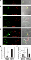
Lasso and LPH1 interact on the cell surface and across intercellular junctions. (A) LPH-51 binds to cell-surface Lasso. NB2a cells expressing Lasso-A were incubated with LPH-51, fixed, and stained with V5 Ab (LPH-51) and with myc Ab (Lasso-A). (B) Soluble Lasso binds to cell-surface LPH1. Cells expressing LPH1-FS were incubated with Lasso-D, fixed, and stained with V5 Ab (LPH1-FS) and with FLAG Ab (Lasso-D). (C and D) Interaction between Lasso and LPH1 on opposite cells. NB2a cells expressing Lasso-A were cocultured with cells expressing a control GPCR (EMR2) (C) or LPH1-FS (D), fixed and stained for Lasso-A (green) and LPH-51 or EMR2 (red). Note that Lasso-A and LPH1-FS concentrate (arrowheads) and overlap (yellow color) at cell contacts. (Scale bars: 20 μm.) (E) Colocalization of Lasso-A with LPH1-FS (but not EMR2) at cell contacts (mean ± SEM; t test; n = 10). (F) Aggregation of Lasso-A--expressing cells with cells expressing LPH1-FS but not LPH2-FS. Gray bars, the number of cell clusters per field of view; black bars, the number of clusters > 100 μm (mean ± SEM; t test; n = 5).
Fig. 4.
Fig. 4.
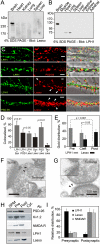
Lasso and LPH1 are expressed on opposite membranes at synaptic junctions. (A and B) Lasso and LPH1 are brain-specific. Detergent extracts from rat tissues were analyzed by immunoblotting with dmAb or PAL1 Ab (typical results; n = 3–7). (C) Lasso and LPH1 are enriched in synapses. Hippocampal neurons (14 d in vitro) were immunostained for LPH1, synapsin, PSD-95, and Lasso, as indicated (n = 5–8 for each Ab). Arrowheads, dendritic spines. (Scale bars: 2 μm.) (D) Colocalization of LPH1 and Lasso with presynaptic and postsynaptic markers and the axonal protein tau in hippocampal cultures (images as in C and Fig. S3; the t tests compare each pair to tau+Lasso). (E) Quantitation of immunoelectron microscopy images (see F and G below and Fig. S4). The total number of gold particles in 517 micrographs was 1,241 for LPH1, 974 for control, and 558 for Lasso. (F and G) Immunoelectron microscopy of LPH1 (F) and Lasso (G) in isolated rat brain synapses. Synaptosomes were incubated with anti-LPH1 Ab A1 (F) or 100 nM LPH-51 (G), counterstained with anti-V5 Ab conjugated with 5 nm immunogold, processed, and imaged by electron microscopy. Synaptic terminals were identified as containing synaptic vesicles and an attached postsynaptic membrane with characteristic intracellular structures. (H) LPH1 is presynaptic and Lasso is postsynaptic in central synapses. Synaptic junction membranes were separated, enriched with presynaptic and postsynaptic membranes (37), and analyzed by immunoblotting with Abs against Lasso, LPH1, NMDAR, Kv1.2, and PSD-95. Note that Lasso distribution is similar to NMDAR but opposite of LPH1. (I) Quantitation of LPH1 and Lasso distribution (mean ± SEM; n = 5).
Fig. 5.
Fig. 5.
LPH1 is recruited to artificial synapses. Cocultures of HEK293 cells expressing Lasso-A (A) or LPH1-FS (B) with primary hippocampal neurons were immunostained for respective tags and for the neuronal postsynaptic protein PSD-95. LPH1, but not Lasso, is recruited (empty arrowheads) to artificial junctions between HEK cells and en passant neuronal dendrites (filled arrowheads), denoted by PSD-95 (n = 5).
Fig. 6.
Fig. 6.
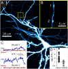
C terminus of Lasso stimulates presynaptic Ca2+ signaling in cultured hippocampal neurons. (A) Fluorescence image of a typical hippocampal neuron loaded with the morphological tracer Alexa Fluor 568 and the Ca2+ dye Fluo-4 (only tracer fluorescence is shown). (B) A fragment of the axon containing two putative presynaptic boutons (magnified from the boxed area in A). (C) Normalized Ca2+ fluorescence responses in boutons 1 and 2, before (red) and 5 min after (blue) application of Lasso-G. (D) Relative change of Ca2+ fluorescence averaged over 15 min after application of Lasso-G (4 cells; 16 boutons) or in control conditions (6 cells; 22 boutons). Red lines represent median responses. Wilcoxon rank tests: control vs. 1, P > 0.8; Lasso-G vs. 1, P < 0.001; Lasso-G vs. control, P < 0.002.

References

    1. Davletov BA, Shamotienko OG, Lelianova VG, Grishin EV, Ushkaryov YA. Isolation and biochemical characterization of a Ca2+-independent α-latrotoxin–binding protein. J Biol Chem. 1996;271:23239–23245. - PubMed
    1. Lelianova VG, et al. The α-latrotoxin receptor latrophilin is a novel member of the secretin family of G protein–coupled receptors. J Biol Chem. 1997;272:21504–21508. - PubMed
    1. Matsushita H, Lelianova VG, Ushkaryov YA. The latrophilin family: Multiply spliced G protein–coupled receptors with differential tissue distribution. FEBS Lett. 1999;443:348–352. - PubMed
    1. Volynski KE, et al. Mutant α-latrotoxin (LTXN4C) does not form pores and causes secretion by receptor stimulation: This action does not require neurexins. J Biol Chem. 2003;278:31058–31066. - PubMed
    1. Volynski KE, et al. Latrophilin fragments behave as independent proteins that associate and signal on binding of LTX(N4C) EMBO J. 2004;23:4423–4433. - PMC - PubMed

Publication types