. 2012 Feb 19;483(7388):176-81.
doi: 10.1038/nature10812.

Piezo proteins are pore-forming subunits of mechanically activated channels

Affiliations

Piezo proteins are pore-forming subunits of mechanically activated channels

Bertrand Coste et al. Nature. .

Abstract

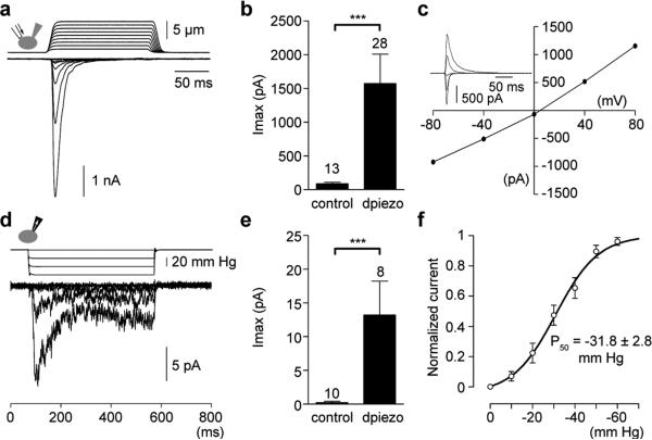
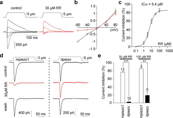
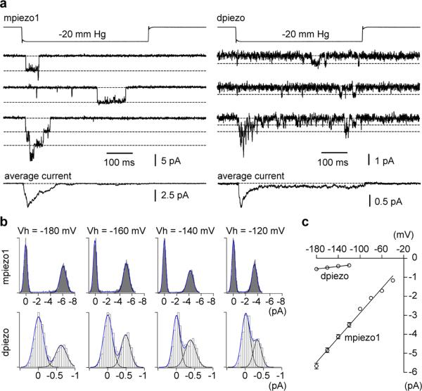
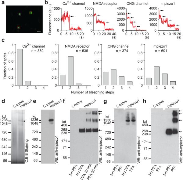
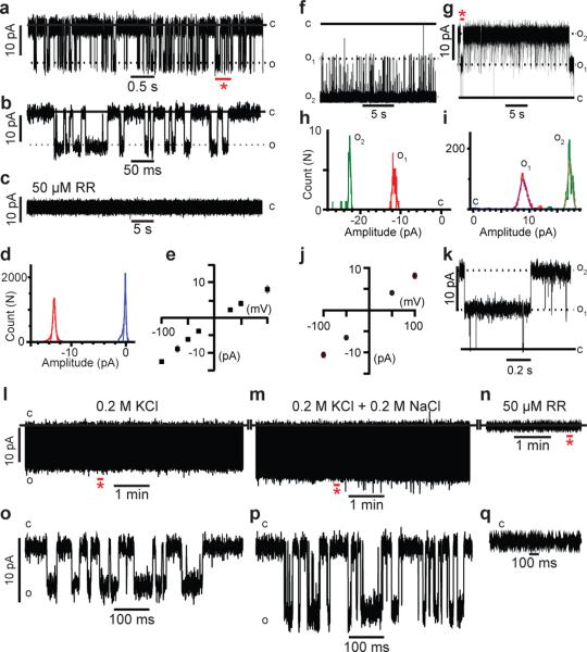
Mechanotransduction has an important role in physiology. Biological processes including sensing touch and sound waves require as-yet-unidentified cation channels that detect pressure. Mouse Piezo1 (MmPiezo1) and MmPiezo2 (also called Fam38a and Fam38b, respectively) induce mechanically activated cationic currents in cells; however, it is unknown whether Piezo proteins are pore-forming ion channels or modulate ion channels. Here we show that Drosophila melanogaster Piezo (DmPiezo, also called CG8486) also induces mechanically activated currents in cells, but through channels with remarkably distinct pore properties including sensitivity to the pore blocker ruthenium red and single channel conductances. MmPiezo1 assembles as a ∼1.2-million-dalton homo-oligomer, with no evidence of other proteins in this complex. Purified MmPiezo1 reconstituted into asymmetric lipid bilayers and liposomes forms ruthenium-red-sensitive ion channels. These data demonstrate that Piezo proteins are an evolutionarily conserved ion channel family involved in mechanotransduction.


Figures

Figure 1
Figure 1. Human cells expressing Drosophila piezo show large mechanically activated currents
MA currents of dpiezo-expressing HE293T cells recorded in the whole-cell (a-c) or cell-attached (d-f) configuration. (a) Representative traces of MA inward currents at -80 mV in dpiezotransfected cells subjected to a series of mechanical steps in 1 μm increments . (b) Average maximal current amplitude of MA inward currents at -80mV. (c) Representative current-voltage relationship of MA currents in dpiezo-transfected cells. (Inset) MA currents evoked at holding potentials ranging from -80 to +80 mV. (d) Representative currents elicited by negative pipette pressure (0 to -60 mmHg, Δ20 mmHg) in dpiezo-transfected cells. (e) Average maximal current amplitude of stretch activated currents at -80mV. (f) Imax normalized current-pressure relationship of stretch activated currents recorded at -80 mV in dpiezo-transfected cells (n = 8 cells) and fitted with a Boltzmann equation. P50 is the average of P50 values determined for individual cells. Bars represent mean ± s.e.m. and the number of cells tested is shown above bars. *** P<0.001, Mann-Whitney test.
Figure 2
Figure 2. Ruthenium red is a channel pore blocker of mpiezo1- but not dpiezo-induced currents
(a) Representative traces of MA currents in mpiezo1-transfected cells evoked at holding potentials ranging from -80 to +80 mV before (left panel) and during perfusion of 30 μM of RR (right panel, red traces). (b) Average current-voltage relationship of MA currents in mpiezo1-transfected cells (n = 7 cells) before (black symbols) and during (red symbols) perfusion of 30 μM RR. Currents were normalized to the value of control current evoked at -80 mV for each individual cell. (c) Concentration-inhibition curve for RR on MA currents evoked at -80 mV in mpiezo1-transfected cells and fitted with a Boltzmann equation. Each data point is the mean ± s.e.m. of 3-13 observations. (d) Representative traces of piezo-dependent MA currents evoked at -80 mV in the presence of RR. (e) Blocking effect of RR on piezo-dependent MA currents evoked at -80 mV. Bars represent mean ± s.e.m. and the number of cells tested is shown above bars. *** P<0.001; ** P<0.01, unpaired t-test.
Figure 3
Figure 3. mpiezo1- and dpiezo-induced stretch-activated channels have different conductances
(a) Representative piezo-dependent stretch-activated channel openings elicited at -180 mV. Bottom traces represent average of 40 individual recording traces. (b) All-point histograms of single channel opening events (average of 10 and 20 individual events for mpiezo1 and dpiezo, respectively) at different holding potentials. (c) Average current-voltage relationships of stretch activated single channels in mpiezo1 and dpiezo transfected cells (n = 7 and 5 cells, respectively; mean ± s.e.m.). Single channel amplitude was determined as the amplitude difference in Gaussian fits as shown in b.
Figure 4
Figure 4. mpiezo1 forms tetramers
(a) Representative image of an acquired sequence showing three selected GFP-mpiezo1 spots in the cell membrane. Levels were adjusted for clarity. Scale bar, 0.8 μm. (b) Representative traces of fluorescence intensities of indicated single GFP-fusion constructs. Black arrows indicate photo-bleaching steps. (c) Histograms of the average number of bleaching steps observed in 10 or more movies from 4 or more oocytes of single fluorescent complexes of indicated constructs. (d,e) Indicated samples purified and separated on native gels and visualized by Coomassie staining (d) or western blotting (e). Asterisk in (d) indicates a strong protein band specifically present in the mpiezo1 sample. (f) Purified mpiezo1-GST proteins treated with or without PFA with the indicated time period, separated on a denaturing gel and detected with the anti-mpiezo1 antibody. Sample purified from cells without transfection served as a negative control. (g, h) mpiezo1-GST-transfected HEK293T cells or untransfected cells treated with or without 0.25% PFA for 10 minutes. The crosslinked mpiezo1-GST proteins were purified and separated on native gel (g) or denaturing gels (h), followed by western blotting. Panels d-h are representatives of at least 3 independent experiments.
Figure 5
Figure 5. mpiezo1 forms ruthenium red sensitive ion channels
(a-e) Reconstitution of purified mpiezo1 into asymmetric lipid bilayers. (a) Representative single channel currents at –100 mV. The section of the recordings indicated by the red asterisk is shown in (b) at a 100-fold higher time resolution. (c) After 35 minutes of recording the channel activity shown in (a), injection of 50 μM RR onto the neutral facing compartment blocks mpiezo1 currents. (d) All-event current amplitude histogram of a 6 minute recording; γ = 124 ± 7 pS. The total number of opening events (N) analyzed was 18,424. (e) Single channel current-voltage (IV) relationship, n=6 experiments. (f-k) Reconstitution of purified mpiezo1 into asolectin proteoliposomes. Representative channel currents recorded at –100 mV (f) and +100 mV (g) in presence of 50 μM RR inside the recording pipette. Two open channels are present in the membrane. The segment of the 15 minute recording shown in (g) indicated by the red asterisk is displayed in (k) at a 25-fold higher time resolution. (h,i) All-event current amplitude histograms from a 30 s (h) and 15 min (i) recordings: γ = 110 ± 10 pS (h) and 80 ± 5 pS (i); N was 9,938 events. (j) Single channel I-V relationship, n=8 experiments. (l-q) Representative single channel currents at -100 mV of purified mpiezo1 reconstituted into asymmetric lipid bilayers in symmetric 0.2 M KCl (l), after addition of 0.2 M NaCl (m) and after addition of 50 μM RR (n). Segments indicated by red asterisks in panels l-n are displayed in panels o-q, respectively. C and O denote the closed and open states.

Comment in

References

    1. Corey DP, Hudspeth AJ. Response latency of vertebrate hair cells. Biophys J. 1979;26:499–506. - PMC - PubMed
    1. McCarter GC, Reichling DB, Levine JD. Mechanical transduction by rat dorsal root ganglion neurons in vitro. Neurosci Lett. 1999;273:179–182. - PubMed
    1. Davis MJ, Donovitz JA, Hood JD. Stretch-activated single-channel and whole cell currents in vascular smooth muscle cells. Am J Physiol. 1992;262:C1083–1088. - PubMed
    1. Praetorius HA, Spring KR. Bending the MDCK cell primary cilium increases intracellular calcium. J Membr Biol. 2001;184:71–79. - PubMed
    1. Chalfie M. Neurosensory mechanotransduction. Nat Rev Mol Cell Biol. 2009;10:44–52. - PubMed

Publication types

MeSH terms

Associated data