. 2012 Feb;24(2):823-38.
doi: 10.1105/tpc.112.095984. Epub 2012 Feb 21.

A MAP kinase kinase interacts with SymRK and regulates nodule organogenesis in Lotus japonicus

Affiliations

A MAP kinase kinase interacts with SymRK and regulates nodule organogenesis in Lotus japonicus

Tao Chen et al. Plant Cell. 2012 Feb.

Abstract

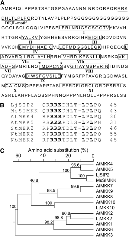
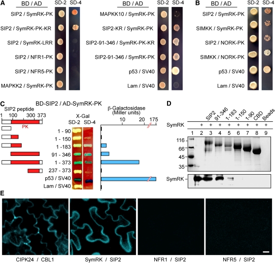
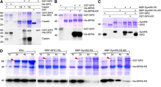
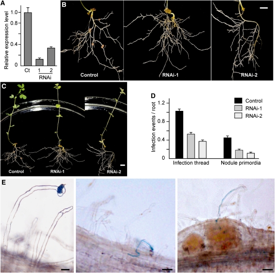
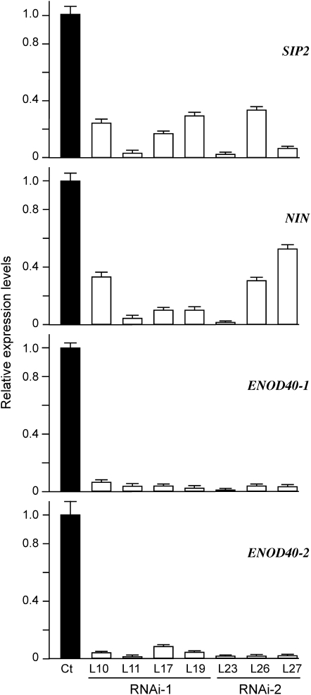
The symbiosis receptor kinase, SymRK, is required for root nodule development. A SymRK-interacting protein (SIP2) was found to form protein complex with SymRK in vitro and in planta. The interaction between SymRK and SIP2 is conserved in legumes. The SIP2 gene was expressed in all Lotus japonicus tissues examined. SIP2 represents a typical plant mitogen-activated protein kinase kinase (MAPKK) and exhibited autophosphorylation and transphosphorylation activities. Recombinant SIP2 protein could phosphorylate casein and the Arabidopsis thaliana MAP kinase MPK6. SymRK and SIP2 could not use one another as a substrate for phosphorylation. Instead, SymRK acted as an inhibitor of SIP2 kinase when MPK6 was used as a substrate, suggesting that SymRK may serve as a negative regulator of the SIP2 signaling pathway. Knockdown expression of SIP2 via RNA interference (RNAi) resulted in drastic reduction of nodules formed in transgenic hairy roots. A significant portion of SIP2 RNAi hairy roots failed to form a nodule. In these roots, the expression levels of SIP2 and three marker genes for infection thread and nodule primordium formation were downregulated drastically, while the expression of two other MAPKK genes were not altered. These observations demonstrate an essential role of SIP2 in the early symbiosis signaling and nodule organogenesis.


Figures

Figure 1.
Figure 1.
SIP2 Is a MAPKK from L. japonicus. (A) Deduced amino acid sequence of SIP2. The conserved DEJL motif is underlined. The catalytic subdomains I to XI of protein kinases are boxed and indicated by Roman numbers. Two putative phosphorylation sites of Thr and Ser are marked with asterisks. The activation loop, which comprises the two phosphorylation residues and is a putative target of upstream MAPKKKs, is underlined twice. (B) The highly conserved DEJL motif of L. japonicus SIP2 and Arabidopsis MAPKKs. It has a consensus of K/R-K/R-K/R-X(1-5)-L/I-X-L/I and is designated for its putative role as the docking site for mammalian ERK and JNK MAPKs or LXL. The highly conserved Leu-X-Leu/Ile (LXL) motif provides a hydrophobic site for binding with MAPKs via hydrophobic interaction. The basic residues (RRK) within the DEJL motif interact with acidic residues of MAPKs via salt bridges. The last residue in each protein is indicated by a number. (C) Phylogenetic tree of L. japonicus SIP2, MAPKK2, and MAPKK10, M. sativa SIMKK, and Arabidopsis MAPKK family members. SIP2 is most closely related to At-MKK4 and At-MKK5. Multiple sequence alignment and neighbor-joining phylogenetic analysis were performed using DNAStar software. Bootstrap values (%) obtained from 1000 trials are given at branch nodes. The alignment used to generate this tree is available as Supplemental Data Set 1 online.
Figure 2.
Figure 2.
Interaction of SIP2 with SymRK in Vitro and in Planta. (A) SIP2 interacts with SymRK in yeast cells. Yeast Y187 cells carrying the Gal4 DNA binding domain (BD) fusion constructs were mated with yeast AH109 cells harboring the Gal4 activation domain (AD) fusion constructs. The diploid cells were selected on SD media lacking Leu and Trp (SD-2), and interaction was assessed according to their ability to grow on selective SD media lacking Leu, Trp, His, and Ade (SD-4) for 5 d. SymRK-LRR, the extracellular LRR region of SymRK; SymRK-PK, the intracellular PK domain of SymRK; NFR1-PK, the intracellular PK domain of NFR1; NFR5-PK, the intracellular PK domain of NFR5; SIP2-KR, a Lys-to-Arg substitution kinase-negative mutant of SIP2; SIP2-91-346, the PK domain of SIP2. Note that SIP2 did not interact with the putative NF receptors (NFR1 and NFR5), and SymRK did not interact with Lj-MAPKK2 and Lj-MKK10. SIP2 did interact with the SymRK ortholog (NORK) from alfalfa, while SymRK also interacted with the SIP2 ortholog (SIMKK) from alfalfa. The interaction between mammalian p53 and SV40 served as a positive control, whereas coexpression of lamin (Lam) and SV40 served as a negative control. (B) Interactions between SymRK and SIP2 orthologs from Lotus and Medicago. Lotus SymRK interacted with the SIP2 ortholog, SIMKK, from alfalfa, and similarly, Lotus SIP2 interacted with the SymRK ortholog, NORK, from alfalfa. The interaction between alfalfa SIMKK and NORK-PK was relatively weak, and the yeast colonies were selected on SD/-Leu/-Trp/-Ade media, followed by selection on SD/-Leu/-Trp/-Ade/-His. (C) The PK domain of SIP2 is responsible for interaction with SymRK. SIP2 and its truncated constructs were expressed as fusion proteins with the GAL4 binding domain (BD) in pGBKT7. SymRK-PK was expressed as a recombinant protein fused with the GAL4 activation domain (AD) in pGADT7. Yeast cells containing both plasmids were grown on SD-Leu-Trp medium (SD-2) containing X-Gal (80 mg/L) and assessed for interactions on SD-Leu-Trp-His-Ade medium (SD-4) containing X-Gal. The strength of interaction was evaluated by assaying β-galactosidase activities in soluble extracts prepared from yeast colonies. (D) In vitro protein pull-down assay for the interaction between SIP2 and SymRK-PK. CBD-tagged SIP2 and four of its truncated products (numbers indicate residues of SIP2) were absorbed to chitin beads and mixed with purified soluble SymRK-PK. After washing, proteins pulled down by the chitin beads were separated via SDS-PAGE and visualized with Coomassie blue dye (top). The same gel was used for immunoblotting with anti-SymRK antibody (bottom). Note that the full-length SIP2 and its PK domain (residues 91 to 346) could pull down SymRK, and the N-terminal half (1 to 183) of SIP2 also could interact with SymRK with a reduced affinity. (E) BiFC on the interaction between SIP2 and SymRK in planta. N. benthamiana leaves were cotransformed with SCFPC155:SymRK-full and SIP2-full:SCFPN173 (SymRK / SIP2). Leaf epidermal cells were observed via fluorescence microscopy. Because the CFP is split into N- and C-terminal halves and fused with different proteins, no cyan fluorescence would be observed if the fusion proteins did not interact. As a positive control, Arabidopsis CIPK24 (CBL-interacting protein kinase 24) and calcineurin B-like 1 (CBL1) were fused with the C-terminal CFPC155 and N-terminal CFPN173, respectively, and coexpressed. Note that no interactions were observed between NFR1 and SIP2, or NFR5 and SIP2 using similar fusion approach. Bar = 20 μm.
Figure 3.
Figure 3.
In Vitro Kinase Activity of SIP2. (A) Protein kinase activity of SIP2. SIP2 was expressed as a GST fusion protein in vector pGEX-6P1. GST alone served as a control. SIP2 was also expressed as a His tag fusion protein in pET28a. Affinity-purified GST, GST-SIP2, and His-SIP2 were incubated with casein or calf intestinal phosphatase–treated casein in the presence of [γ-32P]ATP. The reaction products were analyzed in SDS-PAGE. The gel was stained with Coomassie blue (top), dried, and subjected to autoradiography (bottom). Molecular mass standard (kD) is shown on left side. (B) Phosphorylation of MPK6 MAPK by SIP2. Purified GST-SIP2 was incubated with Arabidopsis MPK6 or its kinase-negative mutant, MPK6-KR, in the presence of [γ-32P]ATP. Kinase reaction products were resolved on SDS-PAGE. The gel was stained with Coomassie blue (top) and subjected to autoradiography (bottom). (C) SIP2 and SymRK did not phosphorylate each other. Because both SIP2 and SymRK could undergo autophosphorylation, their kinase-negative mutants were used as substrates. SIP2 and its kinase-negative mutant were purified on GST beads, whereas SymRK and its kinase-negative mutant were isolated via the MBP tag using chitin beads. In the presence of [γ-32P]ATP, the kinase-negative SymRK-PK-KR was not phosphorylated by SIP2. Similarly, the kinase-negative SIP2-KR was not phosphorylated by SymRK-PK. (D) SymRK inhibited the phosphorylation of MPK6 by SIP2. The kinase-negative MPK6-KR was purified on Ni beads and used as substrate for SIP2 in the presence of [γ-32P]ATP. An increasing amount of SymRK-PK or its kinase-negative SymRK-PK-KR was added to the reaction mix. Increasing amounts of BSA and purified NFR1-PK were used to test the specificity of inhibition. The base amount (1×) of effector proteins (BSA, NFR1, and SymRK) was 1.0 μg per reaction, and the increasing amounts are indicated as relative quantities (2× to 6×). The bands corresponding to the increased amount of added proteins are labeled by arrows. The reaction products were analyzed on SDS-PAGE gels stained with Coomassie blue (top). Autoradiographs show the corresponding phosphorylated MPK6-KR (bottom). [See online article for color version of this figure.]
Figure 4.
Figure 4.
Gene Expression and Subcellular Localization of SIP2. (A) Expression of SIP2 mRNA in L. japonicus. Roots were harvested 1, 2, 4, 6, 8, 10, and 12 d after inoculation with M. loti. Roots treated with water (uninoculated roots) were also harvested at the same time intervals and served as the mock control. Rhizobium-inoculated roots (IR), stems (S), and leaves (L) were harvested 8 d after inoculation, while nodules (N) were collected 42 d after inoculation. Total RNA was isolated and used for real-time PCR to measure the expression levels of the SIP2 mRNA. The ATPase gene (AW719841) was used as an internal control. Error bars represent sd of the experimental values obtained from three technical replicates. (B) Subcellular localization of SIP2 in L. japonicus hairy roots. SIP2 was expressed as a GFP fusion protein (GFP:SIP2) under the control of the CaMV35S promoter in L. japonicus hairy roots induced by A. rhizogenes LBA1334. GFP alone served as a control. Bars = 20 μm. (C) Subcellular localization of GFP-tagged SIP2 in onion cells. SIP2 was expressed as a fusion protein with GFP (GFP:SIP2) under the control of the 35S promoter. Plasmid expressing GFP alone served as a control. The plasmids were delivered to the onion epidermal cells via particle bombardment, and fluorescence images were taken using a confocal laser scanning microscope. Onion epidermal cells expressing GFP:SIP2 were treated with 4% NaCl for 5 min to induce plasmolysis before imaging. Green fluorescence (left) and the corresponding bright-field images (right) were taken using a confocal laser scanning microscope. The overlay images (middle) were produced using Photoshop software. Bars = 50 μm.
Figure 5.
Figure 5.
Expression of SIP2-RNAi in L. japonicus Hairy Roots. (A) Real-time RT-PCR analysis of SIP2 expression levels in the control hairy roots expressing the empty vector (Ct) and representatives of hairy roots expressing the SIP2-RNAi constructs (RNAi-1 and RNAi-2). Error bars represent sd of the experimental values obtained from three technical replicates. (B) The control hairy roots expressing the empty vector and representative hairy roots expressing the SIP2-RNAi constructs (RNAi-1 and RNAi-2). L. japonicus plants were maintained in a nitrogen fertilizer-free environment and root images were taken 4 weeks after Rhizobium infection. Bar = 1 cm. (C) Images of whole plants whose roots were shown in (B). Bar = 1 cm. (D) Numbers of ITs and nodule primordia per root of the control hairy roots and SIP2-RNAi-1 and SIP2-RNAi-2 hairy roots. The data on ITs and nodule primordia were collected from 15 to 20 independent hairy roots in each group of plants. Error bars represent sd of the experimental values. (E) IT formation in SIP2-RNAi hairy roots. ITs were visualized after staining with X-Gal in hairy roots 8 d after infection with M. loti expressing a lacZ construct. The images show the key steps of Rhizobium infection in hairy roots, including bacterial entry and root hair deformation (left), IT growth (middle), and release of rhizobial cells in developing nodule cells (right). Bars = 20 μm in left and middle panels and 50 μm in the right panel.
Figure 6.
Figure 6.
Suppression of Marker Genes Related to IT Formation and Nodule Organogenesis. Total RNA was isolated from the control hairy roots (Ct) expressing the empty vector and representative plants expressing SIP2-RNAi-1 (L10, L11, L17, and L19) and SIP2-RNAi-2 (L23, L26, and L27). Real-time RT-PCR analysis was performed to assess the expression levels of SIP2, NIN, ENOD40-1, and ENOD40-2 genes. Error bars represent sd of the experimental values obtained from three technical replicates.

References

    1. Asai T., Tena G., Plotnikova J., Willmann M.R., Chiu W.L., Gomez-Gomez L., Boller T., Ausubel F.M., Sheen J. (2002). MAP kinase signalling cascade in Arabidopsis innate immunity. Nature 415: 977–983 - PubMed
    1. Bardwell L., Cook J.G., Chang E.C., Cairns B.R., Thorner J. (1996). Signaling in the yeast pheromone response pathway: Specific and high-affinity interaction of the mitogen-activated protein (MAP) kinases Kss1 and Fus3 with the upstream MAP kinase kinase Ste7. Mol. Cell. Biol. 16: 3637–3650 - PMC - PubMed
    1. Beck M., Komis G., Müller J., Menzel D., Samaj J. (2010). Arabidopsis homologs of nucleus- and phragmoplast-localized kinase 2 and 3 and mitogen-activated protein kinase 4 are essential for microtubule organization. Plant Cell 22: 755–771 - PMC - PubMed
    1. Beck M., Komis G., Ziemann A., Menzel D., Samaj J. (2011). Mitogen-activated protein kinase 4 is involved in the regulation of mitotic and cytokinetic microtubule transitions in Arabidopsis thaliana. New Phytol. 189: 1069–1083 - PubMed
    1. Capoen W., Goormachtig S., De Rycke R., Schroeyers K., Holsters M. (2005). SrSymRK, a plant receptor essential for symbiosome formation. Proc. Natl. Acad. Sci. USA 102: 10369–10374 - PMC - PubMed

Publication types

MeSH terms

Substances

LinkOut - more resources