. 2012 Sep;24(9):3559-74.
doi: 10.1105/tpc.112.100511. Epub 2012 Sep 11.

Repeatless and repeat-based centromeres in potato: implications for centromere evolution

Affiliations

Repeatless and repeat-based centromeres in potato: implications for centromere evolution

Zhiyun Gong et al. Plant Cell. 2012 Sep.

Abstract

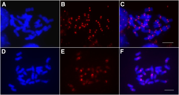
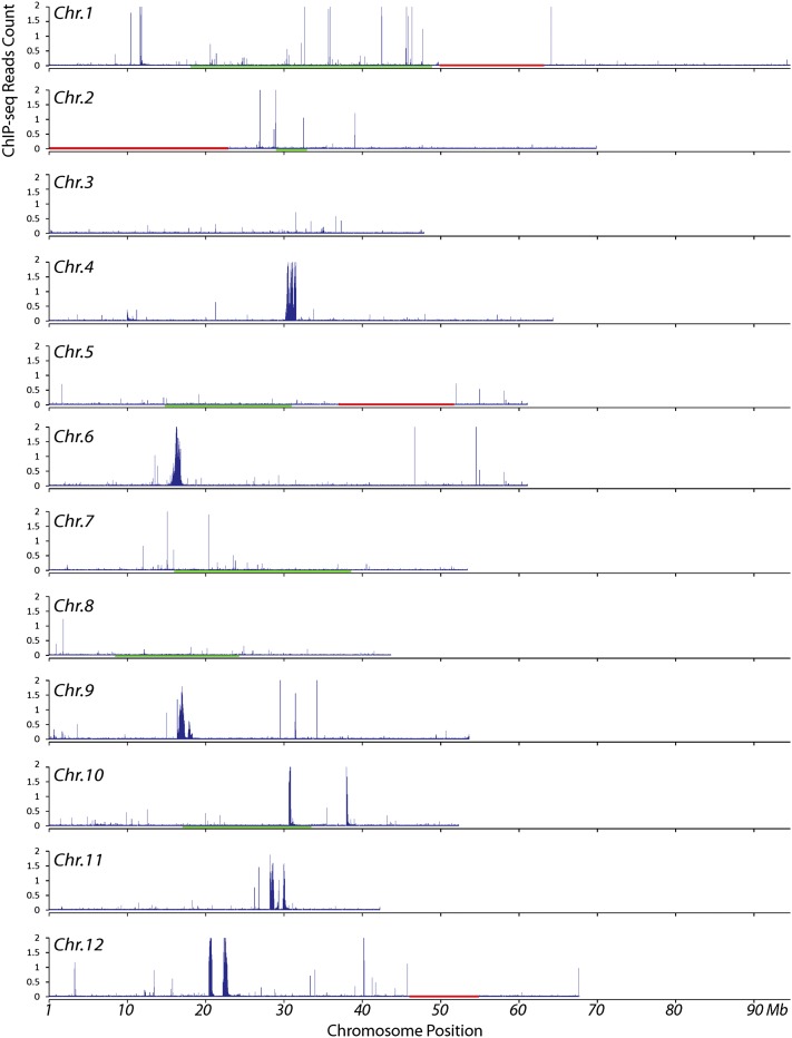
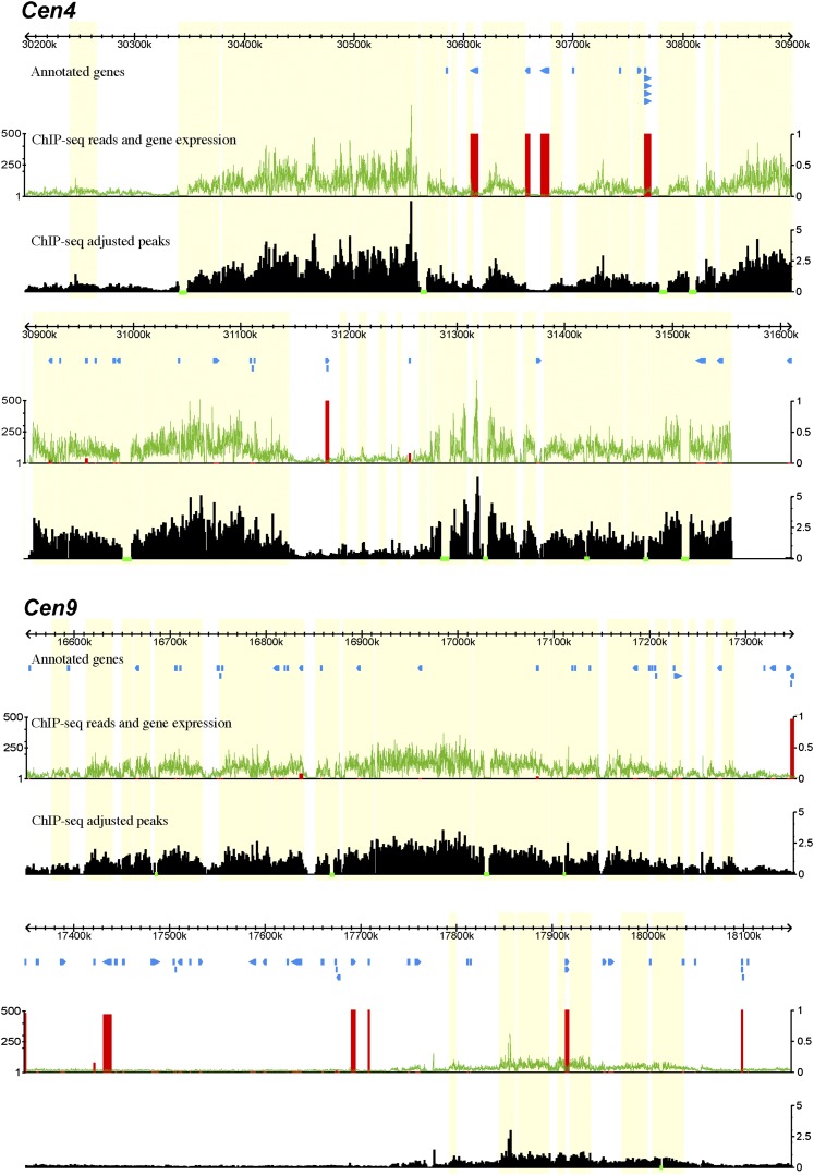
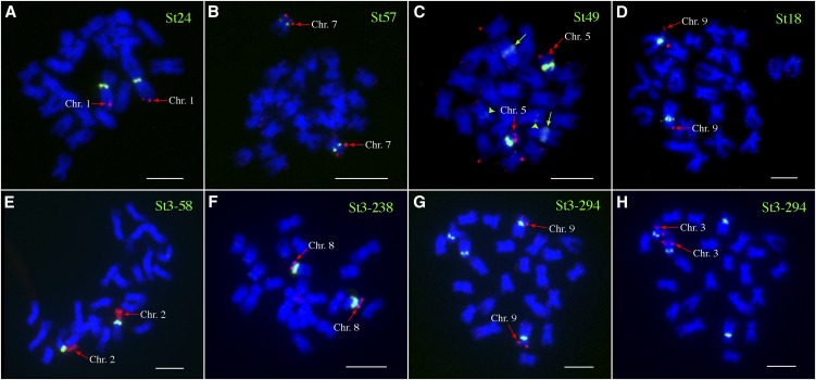
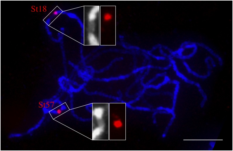
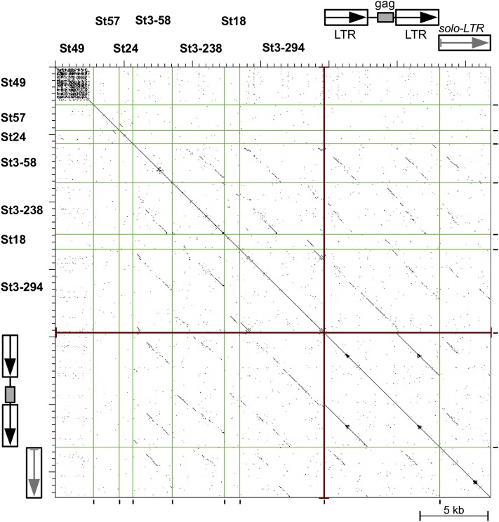
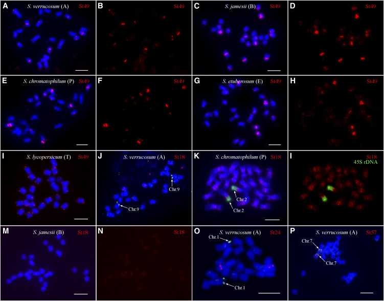
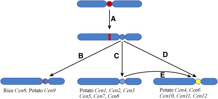
Centromeres in most higher eukaryotes are composed of long arrays of satellite repeats. By contrast, most newly formed centromeres (neocentromeres) do not contain satellite repeats and instead include DNA sequences representative of the genome. An unknown question in centromere evolution is how satellite repeat-based centromeres evolve from neocentromeres. We conducted a genome-wide characterization of sequences associated with CENH3 nucleosomes in potato (Solanum tuberosum). Five potato centromeres (Cen4, Cen6, Cen10, Cen11, and Cen12) consisted primarily of single- or low-copy DNA sequences. No satellite repeats were identified in these five centromeres. At least one transcribed gene was associated with CENH3 nucleosomes. Thus, these five centromeres structurally resemble neocentromeres. By contrast, six potato centromeres (Cen1, Cen2, Cen3, Cen5, Cen7, and Cen8) contained megabase-sized satellite repeat arrays that are unique to individual centromeres. The satellite repeat arrays likely span the entire functional cores of these six centromeres. At least four of the centromeric repeats were amplified from retrotransposon-related sequences and were not detected in Solanum species closely related to potato. The presence of two distinct types of centromeres, coupled with the boom-and-bust cycles of centromeric satellite repeats in Solanum species, suggests that repeat-based centromeres can rapidly evolve from neocentromeres by de novo amplification and insertion of satellite repeats in the CENH3 domains.


Figures

Figure 1.
Figure 1.
Immunofluorescence and ChIP-FISH Using Potato CENH3 Antibodies. (A) Somatic metaphase chromosomes of DM1-3 potato. (B) Immunofluorescence derived from the anti-CENH3 antibodies. (C) Image merged from (A) and (B). (D) Somatic metaphase chromosomes of DM1-3 potato. (E) FISH signals derived from precipitated DNA isolated from ChIP using anti-CENH3 antibodies. (F) Image merged from (D) and (E). Bars = 5 μm.
Figure 2.
Figure 2.
Density of Sequence Reads Derived from ChIP with CENH3 Antibodies along Individual Potato Chromosomes. Read density was represented by the total number of sequence reads in a 10-kb window per base pair mappable region. The x axes show the position on the chromosome. Red horizontal bars mark large physical gaps (>1 Mb) in the pseudomolecules. The red horizontal bar on chromosome 2 represents the unassembled 45S ribosomal gene cluster. The genetic positions of the centromeres of chromosomes 1, 2, 5, 7, 8, and 10 are likely spanned by the green horizontal bars that mark the recombination-suppressed domains on the corresponding linkage maps of these chromosomes (Felcher et al., 2012).
Figure 3.
Figure 3.
Fine Structure of the Functional Cores of Potato Cen4 and Cen9. The top track in each panel shows the sequence coordinates on the respective potato chromosome. The “annotated genes” track illustrates the positions of annotated genes. The green line in the middle track shows the number of sequence reads derived from ChIP with CENH3 antibodies in 100-bp windows. The vertical red bars represent the percentage of the 32 tissues in which the corresponding gene is expressed (FPKM >0) (1 representing expression in all 32 tissues). The bottom track shows the density of reads in 100-bp windows, adjusted by the length of mappable regions. The horizontal green bars in this track mark the sequencing gap/nonmappable regions. Each green bar region is assigned to an adjacent CENH3 subdomain. All CENH3 subdomains are shaded in yellow.
Figure 4.
Figure 4.
RT-PCR Analysis of Two Potato Genes Located in the CENH3 Subdomains. All RT-PCR experiments were conducted using young leaves of DM1-3. (A) Cycle-limited RT-PCR analysis of PGSC0003DMG400012074 (2074) and PGSC0003DMG400039086 (primer pair 9086_1; see Supplemental Table 3 online). 1, DM1-3 cDNA; 2, DM1-3 genomic DNA; Tn, technical negative control (water); L, DNA ladder. PGSC0003DMG400043879 (3879) is an untranscribed gene located in Cen12 and is used as a negative control. The expression of the Urease gene (Ure) and Actin gene (Actin) was used for comparison. (B) Gel electrophoresis analysis of the expression of the genes as in (A) after 40 PCR cycles. No amplicons were obtained for PGSC0003DMG400039086 (both primer pair 9086_1 and primer pair 9086_2) and PGSC0003DMG400043879 (3879).
Figure 5.
Figure 5.
FISH Mapping of Centromeric Repeats in DM1-3 Potato. (A) Repeat St24 was mapped to Cen1 together with BAC clone 96H03, which is specific to 1L. (B) Repeat St57 was mapped to Cen7 together with BAC clone 186I02, which is specific to 7S. (C) Repeat FISH St49 was mapped to Cen5 together with BAC clone 44A21, which is specific to 5S. Two green arrows point to the second signals that are much weaker than the Cen5-specific signals. The two green arrowheads point to the third signals that were very weak but consistently observed. (D) Repeat St18 was mapped to Cen9 together with BAC clone 135I22, which is specific to 9S. (E) Repeat St3-58 was mapped to Cen2 together with the 45S rDNA probe, which is specific to 2S. (F) Repeat St3-238 was mapped to Cen8 together with BAC clone 122L16, which is specific to 8S. (G) Repeat St3-294 was mapped to the centromeres of two pairs of chromosomes. The first pair of chromosomes were identified to be chromosome 9 using BAC clone 135I22, which is specific to 9S. (H) The second pair of chromosomes hybridized to St3-294 were identified to be chromosomes 3 using BAC clone 79E02, which is specific to 3L. Bars = 5 μm.
Figure 6.
Figure 6.
Locations of St18 and St57 on the Pachytene Chromosomes of DM1-3 Potato. St18 is located at the edge of the primary constriction of chromosome 9. FISH signal from St57 is almost completely located within the primary constriction of chromosome 7. Chromosomes were stained by DAPI. Note: The primary constriction of the pachytene chromosomes can be readily identified based on their distinctly lower level of staining compared with the brightly stained pericentromeric heterochromatin. Bar = 10 μm.
Figure 7.
Figure 7.
Dot-Plot Similarity Comparison of Potato Centromeric Satellite Sequences and Selected Retrotransposon-Like Sequences. Individual sequences are separated by vertical lines and their similarities exceeding 60% over a 100-bp sliding window are displayed as black dots or diagonal lines. The retrotransposon-like sequences with similarities to St3 and St18 satellites are represented by nonautonomous LTR retroelement (NA-RE) and related solo-LTR sequence, both identified in potato BAC clone BA251C18 (GenBank accession number GU906238, positions 23518-24812/28493-35636 and 24813-28492, respectively). [See online article for color version of this figure.]
Figure 8.
Figure 8.
Phylogenetic Relationships of the Solanum Species Used in Evolutionary Study of the Potato Centromere-Specific Satellite Repeats. The bootstrap values were based on plastid DNA analyses of Spooner et al. (1993), Spooner and Castillo (1997), and Castillo and Spooner (1997).
Figure 9.
Figure 9.
FISH Mapping of Potato Centromere-Specific Satellite Repeats in Different Solanum Species. (A) FISH of St49 on metaphase chromosomes of S. verrucosum. (B) Digitally separated FISH signals from (A). (C) FISH of St49 on metaphase chromosomes of S. jamesii. (D) Digitally separated FISH signals from (C). (E) FISH of St49 on metaphase chromosomes of S. chromatophilum. (F) Digitally separated FISH signals from (E). (G) FISH of St49 on metaphase chromosomes of S. etuberosum. (H) Digitally separated FISH signals from (G). (I) FISH of St49 on metaphase chromosomes of tomato (S. lycopersicum). (J) FISH of St18 on metaphase chromosomes of S. verrucosum. St18 is not located on chromosome 9, which is identified by BAC 135I22 (arrows). (K) FISH of St18 on metaphase chromosomes of S. chromatophilum. Chromosome 2 is identified by the FISH signals from 45S rDNA (arrows). (L) Digitally separated FISH signals from (K). (M) FISH of St18 on metaphase chromosomes of S. jamesii. (N) Digitally separated FISH signals from (M). (O) FISH of St24 on metaphase chromosomes of S. verrucosum. St24 is not located on chromosome 1, which is identified by BAC 96H03 (arrows). (P) FISH of St57 on metaphase chromosomes of S. verrucosum. St57 is colocalized on chromosome 7, which is identified by BAC 186I02 (arrows). Letters in parentheses represent the genome of the Solanum species. Bars = 5 μm.
Figure 10.
Figure 10.
A Model of Centromere Evolution. (A) A neocentromere activation event may result in the repositioning of the centromere. (B) The evolutionarily new centromere acquired a satellite repeat array during evolution. The satellite repeat may be derived from other centromeres, such as rice Cen8, or a new repeat, such as potato Cen9. The satellite repeat array in the evolutionarily new centromere may expand and eventually occupy the entire centromere. (C) The evolutionarily new centromere may survive for several million years without satellite repeat invasion. Such evolutionarily new centromeres will slowly evolve by accumulating DNA mutations and transposable elements (white lines). (D) and (E) A de novo DNA amplification of a satellite repeat, possibly based on an eccDNA-mediated mechanism, and insertion of the repeat (yellow) in the CENH3 domain can turn an evolutionarily new centromere into a repeat-based centromere.

Comment in

References

    1. Allshire R.C., Nimmo E.R., Ekwall K., Javerzat J.P., Cranston G. (1995). Mutations derepressing silent centromeric domains in fission yeast disrupt chromosome segregation. Genes Dev. 9: 218–233 - PubMed
    1. Black B.E., Brock M.A., Bédard S., Woods V.L., Jr, Cleveland D.W. (2007). An epigenetic mark generated by the incorporation of CENP-A into centromeric nucleosomes. Proc. Natl. Acad. Sci. USA 104: 5008–5013 - PMC - PubMed
    1. Cardone M.F., et al. (2006). Independent centromere formation in a capricious, gene-free domain of chromosome 13q21 in Old World monkeys and pigs. Genome Biol. 7: R91. - PMC - PubMed
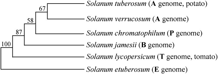
    1. Castillo R.O., Spooner D.M. (1997). Phylogenetic relationships of wild potatoes, Solanum series Conicibaccata (sect. Petota). Syst. Bot. 22: 45–83
    1. Cheng Z.K., Buell C.R., Wing R.A., Jiang J. (2002). Resolution of fluorescence in-situ hybridization mapping on rice mitotic prometaphase chromosomes, meiotic pachytene chromosomes and extended DNA fibers. Chromosome Res. 10: 379–387 - PubMed

Publication types

LinkOut - more resources