. 2013 Jun;87(11):6346-58.
doi: 10.1128/JVI.00416-13. Epub 2013 Mar 27.

Identification and characterization of genetically divergent members of the newly established family Mesoniviridae

Affiliations

Identification and characterization of genetically divergent members of the newly established family Mesoniviridae

Florian Zirkel et al. J Virol. 2013 Jun.

Abstract

The recently established family Mesoniviridae (order Nidovirales) contains a single species represented by two closely related viruses, Cavally virus (CavV) and Nam Dinh virus (NDiV), which were isolated from mosquitoes collected in Côte d'Ivoire and Vietnam, respectively. They represent the first nidoviruses to be discovered in insects. Here, we report the molecular characterization of four novel mesoniviruses, Hana virus, Méno virus, Nsé virus, and Moumo virus, all of which were identified in a geographical region in Côte d'Ivoire with high CavV prevalence. The viruses were found with prevalences between 0.5 and 2.8%, and genome sequence analyses and phylogenetic studies suggest that they represent at least three novel species. Electron microscopy revealed prominent club-shaped surface projections protruding from spherical, enveloped virions of about 120 nm. Northern blot data show that the four mesoniviruses analyzed in this study produce two major 3'-coterminal subgenomic mRNAs containing two types of 5' leader sequences resulting from the use of different pairs of leader and body transcription-regulating sequences that are conserved among mesoniviruses. Protein sequencing, mass spectroscopy, and Western blot data show that mesonivirus particles contain eight major structural protein species, including the putative nucleocapsid protein (25 kDa), differentially glycosylated forms of the putative membrane protein (20, 19, 18, and 17 kDa), and the putative spike (S) protein (77 kDa), which is proteolytically cleaved at a conserved site to produce S protein subunits of 23 and 57 kDa. The data provide fundamental new insight into common and distinguishing biological properties of members of this newly identified virus family.


Figures

Fig 1
Fig 1
Growth and morphology of mesoniviruses. (A) C6/36 cells were infected with CavV, HanaV, NséV, and MénoV, respectively, at an MOI of 0.001 TCID50 per cell, and the numbers of genome copies per milliliter of cell culture supernatant were measured by specific RT-PCR at the indicated time points postinfection. (B) Negative-staining electron micrograph of NséV (isolate NséV/F7/CI/2004) sedimented by ultracentrifugation through a 36% sucrose cushion.
Fig 2
Fig 2
Mesonivirus genome organization. Shown is a schematic view of CavV, HanaV, MénoV, and NséV genomes. Open reading frames are shown by boxes, with nucleotide positions indicated. Conserved functional domains encoded by the replicase gene (ORF1a and -1b) are indicated by gray boxes. ORF2a is shown in light blue, ORF2b in green, and ORF3a and -3b in yellow. Hydrophobic (putative transmembrane) regions are marked in blue, and predicted signal peptides are shown in red. The light-gray boxes symbolize several small, nonconserved ORFs in the 3′-terminal regions of the four mesonivirus genomes. The solid gray box symbolizes ORF4, while other small ORFs are indicated by open gray boxes.
Fig 3
Fig 3
Phylogenetic relationships of mesoniviruses. Shown is a maximum-likelihood phylogenetic tree of mesoniviruses and representative nidoviruses based on amino acid sequences of the conserved pp1ab domains RdRp, 3CLpro, Z, Hel, and the putative N and S proteins. Analyses were performed using the BLOSUM62 substitution model without optimization and with an estimated proportion of invariable sites and an estimated gamma distribution parameter with 1,000 bootstrap replicates.
Fig 4
Fig 4
Mesonivirus genomic and subgenomic RNA synthesis. Northern blot analysis of viral RNA isolated from infected C6/36 cells. Digoxigenin (DIG)-labeled probes specific for the 3′ ends of CavV, HanaV, MénoV, and NséV, respectively, were used for detection. RNA from noninfected C6/36 cells was used as a control. A DIG-labeled RNA was used as a size marker (M), with sizes given in nucleotides on the right and sizes of mRNAs given in kilobases on the left. For the detection of viral genomic RNA, a longer exposure time was used (shown at the top). The positions of genome RNA and the two major sgRNAs (mRNA2 and mRNA3) are indicated on the left.
Fig 5
Fig 5
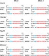
Mesonivirus TRSs. Shown is an overview of TRS core sequences and leader-body fusion sites determined in this study for mRNA2 and -3 of CavV, HanaV, MénoV, and NséV, respectively. The actual fusion sites are shown in blue and the conserved TRS core sequences in red. Production of the two sgRNAs detected in mesonivirus-infected cells involves (i) two different transcription-regulating sequences (TRS1 and -2) and (ii) two different fusion sites of leader and body sequences. As a result, the two major sgRNAs contain 5′ leader sequences of different lengths (∼120 versus ∼20 nucleotides). The genomic positions of leader and body TRSs are indicated.
Fig 6
Fig 6
Mesonivirus structural proteins. (A) SDS-PAGE analysis of the protein content of virus particles purified from cell culture supernatants of infected cells by gradient ultracentrifugation. The proteins were analyzed either directly or after deglycosylation using PNGase F (− and +, respectively). Proteins were stained with Coomassie blue R-250. (B) The identities of CavV virion proteins were analyzed by Edman degradation and MALDI-TOF mass spectroscopy. Sequences identified by Edman degradation are shown in red, and MALDI-TOF sequence coverage is shown in yellow. Specific ORFs shown to encode the respective proteins are given on the right, together with the observed molecular masses of the proteins. Predicted N-linked glycosylation sites in the S and M proteins are indicated by brown triangles. The approximate positions of epitopes recognized by the antibodies used in this study are illustrated by green boxes.
Fig 7
Fig 7
Western blot analysis of mesonivirus proteins. Shown is Western blot analysis of viral proteins present in CavV, HanaV, MénoV, and NséV virions purified by gradient ultracentrifugation. The proteins were analyzed either directly (−) or after deglycosylation using PNGase F (+) and detected using polyclonal rabbit anti-CavV/NséV serum (A), rabbit anti-CavV N serum (B), rabbit anti-CavV S serum (C), and rabbit anti-CavV M serum (D). In panels A and C, proteins shown in the upper blot were separated by SDS-PAGE on a 12% polyacrylamide gel and those in the lower blot on a 14% polyacrylamide gel.

References

    1. Junglen S, Kurth A, Kuehl H, Quan PL, Ellerbrok H, Pauli G, Nitsche A, Nunn C, Rich SM, Lipkin WI, Briese T, Leendertz FH. 2009. Examining landscape factors influencing relative distribution of mosquito genera and frequency of virus infection. Ecohealth 6:239–249 - PMC - PubMed
    1. Zirkel F, Kurth A, Quan PL, Briese T, Ellerbrok H, Pauli G, Leendertz FH, Lipkin WI, Ziebuhr J, Drosten C, Junglen S. 2011. An insect nidovirus emerging from a primary tropical rainforest. mBio 2:e00077–00011. doi:10.1128/mBio.00077-11 - DOI - PMC - PubMed
    1. Nga PT, del Carmen Parquet M, Lauber C, Parida M, Nabeshima T, Yu F, Thuy NT, Inoue S, Ito T, Okamoto K, Ichinose A, Snijder EJ, Morita K, Gorbalenya AE. 2011. Discovery of the first insect nidovirus, a missing evolutionary link in the emergence of the largest RNA virus genomes. PLoS Pathog. 7:e1002215 doi:10.1371/journal.ppat.1002215 - DOI - PMC - PubMed
    1. Lauber C, Ziebuhr J, Junglen S, Drosten C, Zirkel F, Nga PT, Morita K, Snijder EJ, Gorbalenya AE. 2012. Mesoniviridae: a proposed new family in the order Nidovirales formed by a single species of mosquito-borne viruses. Arch. Virol. 157:1623–1628 - PMC - PubMed
    1. Ziebuhr J. 2006. The coronavirus replicase: insights into a sophisticated enzyme machinery. Adv. Exp. Med. Biol. 581:3–11 - PMC - PubMed

Publication types

Substances

Associated data

LinkOut - more resources