. 2013 Jan 2;110(1):193-8.
doi: 10.1073/pnas.1209657109. Epub 2013 Jan 2.

Stable transgenesis in the marine annelid Platynereis dumerilii sheds new light on photoreceptor evolution

Affiliations

Stable transgenesis in the marine annelid Platynereis dumerilii sheds new light on photoreceptor evolution

Benjamin Backfisch et al. Proc Natl Acad Sci U S A. .

Abstract

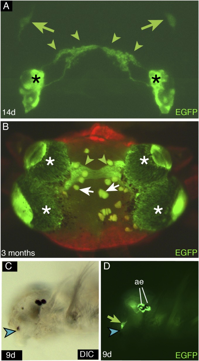
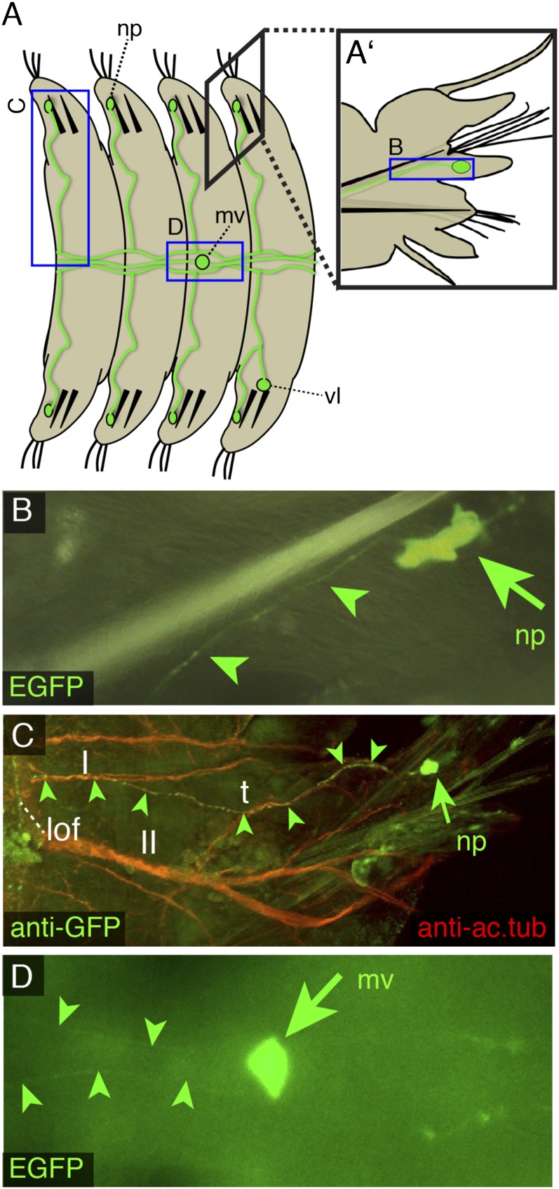
Research in eye evolution has mostly focused on eyes residing in the head. In contrast, noncephalic light sensors are far less understood and rather regarded as evolutionary innovations. We established stable transgenesis in the annelid Platynereis, a reference species for evolutionary and developmental comparisons. EGFP controlled by cis-regulatory elements of r-opsin, a characteristic marker for rhabdomeric photoreceptors, faithfully recapitulates known r-opsin expression in the adult eyes, and marks a pair of pigment-associated frontolateral eyelets in the brain. Unexpectedly, transgenic animals revealed an additional series of photoreceptors in the ventral nerve cord as well as photoreceptors that are located in each pair of the segmental dorsal appendages (notopodia) and project into the ventral nerve cord. Consistent with a photosensory function of these noncephalic cells, decapitated animals display a clear photoavoidance response. Molecular analysis of the receptors suggests that they differentiate independent of pax6, a gene involved in early eye development of many metazoans, and that the ventral cells may share origins with the Hesse organs in the amphioxus neural tube. Finally, expression analysis of opn4×-2 and opn4m-2, two zebrafish orthologs of Platynereis r-opsin, reveals that these genes share expression in the neuromasts, known mechanoreceptors of the lateral line peripheral nervous system. Together, this establishes that noncephalic photoreceptors are more widespread than assumed, and may even reflect more ancient aspects of sensory systems. Our study marks significant advance for the understanding of photoreceptor cell (PRC) evolution and development and for Platynereis as a functional lophotrochozoan model system.


Conflict of interest statement

The authors declare no conflict of interest.

Figures

Fig. 1.
Fig. 1.
Labeling of cephalic rhabdomeric photoreceptor cells (PRCs) by the stable pMos{rops::egfp}vbci2 strain. (A) At 14 d of development, EGFP demarcates the adult eye photoreceptors (asterisks) and their neuronal projections (green arrowheads) as well as a newly identified set of frontolateral eyelets (green arrows). (B) EGFP expression in adult eye PRCs (asterisks) and their projections (green arrowheads) in a mature transgenic animal after spawning. White arrows indicate autofluorescent iridophore pigments. (C and D) The frontolateral eyelet PRCs (green arrow) are frequently found to be associated with pigment cells (blue arrowhead) (A) dorsal view, anterior to the top; (B) frontal view, dorsal to the top; (C and D) lateral views, anterior to the left. ae, adult eyes.
Fig. 2.
Fig. 2.
The pMos{rops::egfp}vbci2 strain reveals a complex set of unpigmented noncephalic photoreceptors. (A and A′) Schematic drawing of four segments of the ventral Platynereis trunk (A) and a view onto a parapodium (A′). Rectangles and labels indicate regions and orientation of A′ and BD; green circles indicate location of noncephalic PRCs, green lines demarcate typical projection trajectories. (BD) Representative examples of noncephalic PRCs (green) visualized by EGFP fluorescence in living animals (B and D) or by anti-GFP immunohistochemistry (C); arrows indicate cell bodies and arrowheads projections. (B and C) Notopodial cell and its projection along nerves II (II) and I (I) into a longitudinal fiber (lof) in the ventral nerve cord; t demarcates turning point away from nerve II; nervous system highlighted by antiacetylated tubulin staining (red). (D) Midventral cell in the ventral nerve cord. (A and D) Ventral views, anterior to the left; (A′ and B) frontal views dorsal to the top; (C) ventral view, anterior to the top. np, notopodial cell; mv, midventral cell; vl, ventrolateral cell.
Fig. 3.
Fig. 3.
The Platynereis trunk shows an autonomous photoavoidance response. (A, C, and D) Schematic outline of light avoidance assay. Decapitated premature worms adapted to dim light were either left in the same conditions (dark) or received a bright light stimulus (light) close to their tail tip (area circled in C). Thirty seconds after starting the experiment, the distance (d) that the illuminated segments had moved was determined. (B) Results of measurements on 20 animals. ****P < 0.0001 in a two-tailed paired t test. Error bars indicate SEM. (Scale bar in C and D, 1 mm.)
Fig. 4.
Fig. 4.
Molecular specification of Platynereis noncephalic photoreceptors and presence of r-opsin orthologs in the zebrafish neuromasts. (AI) Codetection of Platynereis r-opsin (red arrowheads) and indicated transcripts (purple arrowheads) by double in situ hybridization. Coexpression is indicated by black arrows. (AC) Expression in the adult ventral trunk. (DG) Expression in 8-d tail regenerates (trunk and parapodia). (H and I) Expression in adult parapodia. (JL) Expression of opn4x-2 and opn4m-2 in the zebrafish neuromast. (J) Live image of neuromast cells (green arrowheads) in the brn3c::egfp strain 6 d after fertilization; (K and L) corresponding labeling of neuromast cells (purple arrowheads) with the opn4x-2 and opn4m-2 riboprobes. (AG) Ventral views, anterior to the left; (H and I) frontal views, dorsal to the top. (JL) Lateral views, anterior to the left. See text for details.

References

    1. Salvini-Plawen L, Mayr E. On the evolution of photoreceptors and eyes. Evol Biol. 1977;10:207–263.
    1. Dzik J. Early Cambrian lobopodian sclerites and associated fossils from Kazakhstan. Palaeontology. 2003;46(1):93–112.
    1. Purschke G, Ding Z, Müller MC. Ultrastructural differences as a taxonomic marker: The segmental ocelli of Polyophthalmus pictus and Polyophthalmus qingdaoensis sp.n. (Polychaeta, Opheliidae) Zoomorphology. 1995;115(4):229–241.
    1. Hesse R. Untersuchungen über die Organe der Lichtempfindung bei niederen Thieren. V. Die Augen der polychäten Anneliden [Studies on the light sensation in lower animals. V. The eyes of polychaete annelids] Z Wiss Zool. 1899;65:446–516. German.
    1. Xiang Y, et al. Light-avoidance-mediating photoreceptors tile the Drosophila larval body wall. Nature. 2010;468(7326):921–926. - PMC - PubMed

Publication types

Associated data