

Phylogenetic analysis reveals a high prevalence of Sporothrix brasiliensis in feline sporotrichosis outbreaks
- PMID: 23818999
- PMCID: PMC3688539
- DOI: 10.1371/journal.pntd.0002281
Phylogenetic analysis reveals a high prevalence of Sporothrix brasiliensis in feline sporotrichosis outbreaks
Abstract
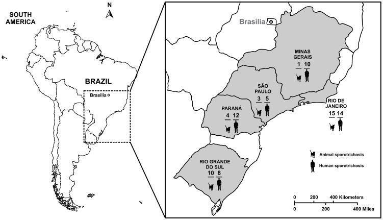
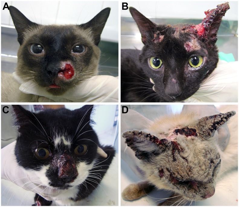
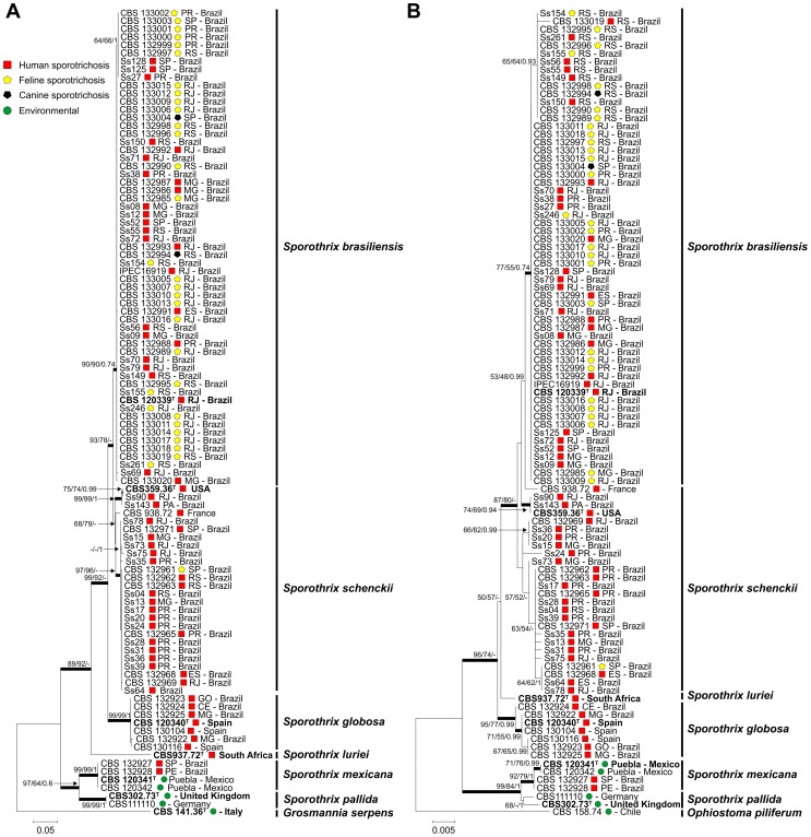
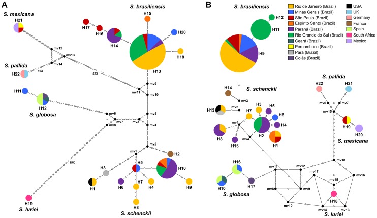
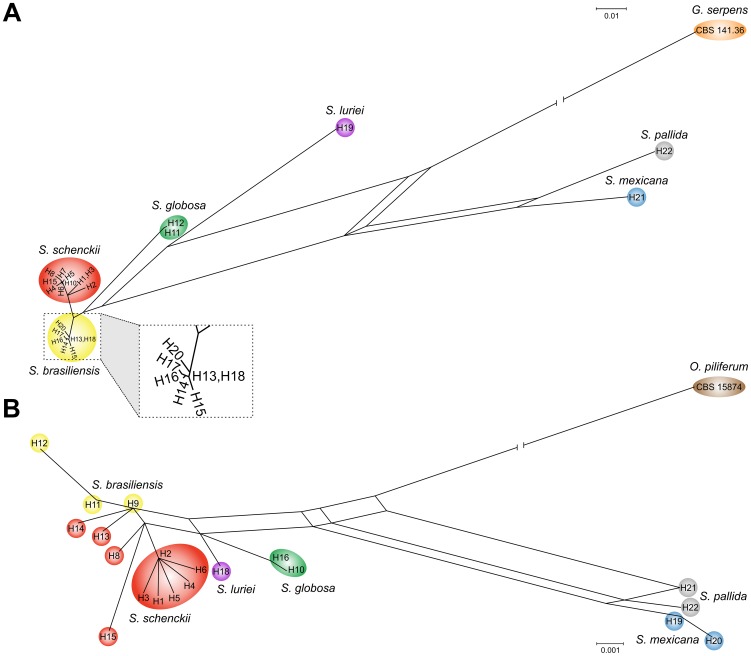
Sporothrix schenckii, previously assumed to be the sole agent of human and animal sporotrichosis, is in fact a species complex. Recently recognized taxa include S. brasiliensis, S. globosa, S. mexicana, and S. luriei, in addition to S. schenckii sensu stricto. Over the last decades, large epidemics of sporotrichosis occurred in Brazil due to zoonotic transmission, and cats were pointed out as key susceptible hosts. In order to understand the eco-epidemiology of feline sporotrichosis and its role in human sporotrichosis a survey was conducted among symptomatic cats. Prevalence and phylogenetic relationships among feline Sporothrix species were investigated by reconstructing their phylogenetic origin using the calmodulin (CAL) and the translation elongation factor-1 alpha (EF1α) loci in strains originated from Rio de Janeiro (RJ, n = 15), Rio Grande do Sul (RS, n = 10), Paraná (PR, n = 4), São Paulo (SP, n =3) and Minas Gerais (MG, n = 1). Our results showed that S. brasiliensis is highly prevalent among cats (96.9%) with sporotrichosis, while S. schenckii was identified only once. The genotype of Sporothrix from cats was found identical to S. brasiliensis from human sources confirming that the disease is transmitted by cats. Sporothrix brasiliensis presented low genetic diversity compared to its sister taxon S. schenckii. No evidence of recombination in S. brasiliensis was found by split decomposition or PHI-test analysis, suggesting that S. brasiliensis is a clonal species. Strains recovered in states SP, MG and PR share the genotype of the RJ outbreak, different from the RS clone. The occurrence of separate genotypes among strains indicated that the Brazilian S. brasiliensis epidemic has at least two distinct sources. We suggest that cats represent a major host and the main source of cat and human S. brasiliensis infections in Brazil.
Conflict of interest statement
The authors have declared that no competing interests exist.
Figures
References
-
- Lutz A, Splendore A (1907) On a mycosis observed in men and mice: Contribution to the knowledge of the so-called sporotrichosis. Revista Médica de São Paulo 21: 443–450 [in Portuguese].
-
- Pereira SA, Menezes RC, Gremião IDF, Silva JN, de O. Honse C, et al. (2011) Sensitivity of cytopathological examination in the diagnosis of feline sporotrichosis. J Feline Med Surg 13: 220–223 doi: 10.1016/j.jfms.2010.10.007 - DOI - PMC - PubMed
-
- Schubach A, Schubach TM, Barros MB, Wanke B (2005) Cat-transmitted sporotrichosis, Rio de Janeiro, Brazil. Emerg Infect Dis 11: 1952–1954 doi: 10.3201/eid1112.040891 - DOI - PMC - PubMed
-
- Schubach TM, Schubach A, Okamoto T, Barros MB, Figueiredo FB, et al. (2004) Evaluation of an epidemic of sporotrichosis in cats: 347 cases (1998–2001). J Am Vet Med Assoc 224: 1623–1629 doi: 10.2460/javma.2004.224.1623 - DOI - PubMed
-
- Mackinnon JE, Conti-Díaz IA, Gezuele E, Civila E, Da Luz S (1969) Isolation of Sporothrix schenckii from nature and considerations on its pathogenicity and ecology. Sabouraudia 7: 38–45 doi:10.1080/00362177085190071 - DOI - PubMed
Publication types
MeSH terms
Substances
Associated data
- Actions
- Actions
- Actions
- Actions
- Actions
- Actions
- Actions
- Actions
- Actions
- Actions
- Actions
- Actions
- Actions
- Actions
- Actions
- Actions
- Actions
- Actions
- Actions
- Actions
- Actions
- Actions
- Actions
- Actions
- Actions
- Actions
- Actions
- Actions
- Actions
- Actions
- Actions
- Actions
- Actions
- Actions
- Actions
- Actions
- Actions
- Actions
- Actions
- Actions
- Actions
- Actions
- Actions
- Actions
- Actions
- Actions
- Actions
- Actions
- Actions
- Actions
- Actions
- Actions
- Actions
- Actions
- Actions
- Actions
- Actions
- Actions
- Actions
- Actions
- Actions
- Actions
- Actions
- Actions
- Actions
- Actions
- Actions
- Actions
- Actions
- Actions
- Actions
- Actions
- Actions
- Actions
- Actions
- Actions
- Actions
- Actions
- Actions
- Actions
- Actions
- Actions
- Actions
- Actions
- Actions
- Actions
- Actions
- Actions
- Actions
- Actions
- Actions
- Actions
- Actions
- Actions
- Actions
- Actions
- Actions
- Actions
- Actions
LinkOut - more resources
Full Text Sources
Other Literature Sources
Research Materials
Miscellaneous
