. 2013 Oct;51(10):3338-57.
doi: 10.1128/JCM.01465-13. Epub 2013 Aug 7.

Molecular characterization of reptile pathogens currently known as members of the chrysosporium anamorph of Nannizziopsis vriesii complex and relationship with some human-associated isolates

Affiliations

Molecular characterization of reptile pathogens currently known as members of the chrysosporium anamorph of Nannizziopsis vriesii complex and relationship with some human-associated isolates

Lynne Sigler et al. J Clin Microbiol. 2013 Oct.

Abstract

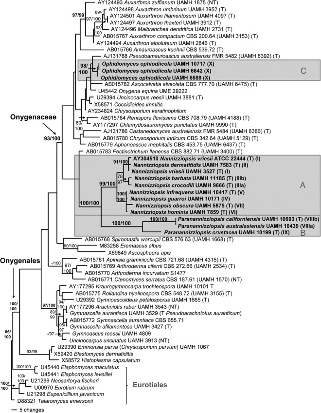
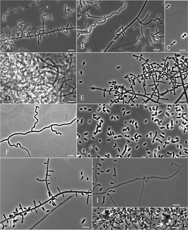
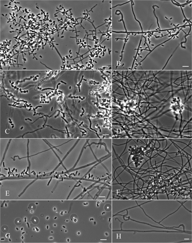
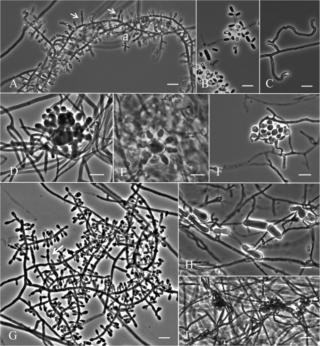
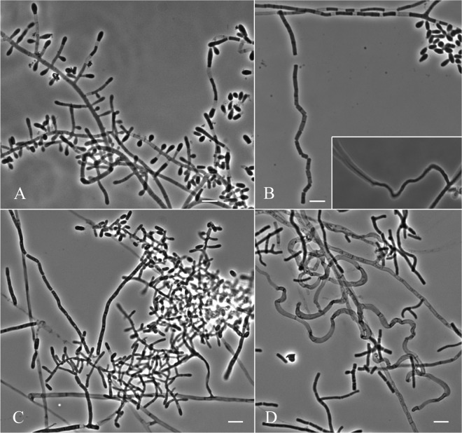
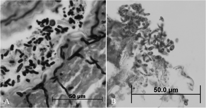
In recent years, the Chrysosporium anamorph of Nannizziopsis vriesii (CANV), Chrysosporium guarroi, Chrysosporium ophiodiicola, and Chrysosporium species have been reported as the causes of dermal or deep lesions in reptiles. These infections are contagious and often fatal and affect both captive and wild animals. Forty-nine CANV isolates from reptiles and six isolates from human sources were compared with N. vriesii based on their cultural characteristics and DNA sequence data. Analyses of the sequences of the internal transcribed spacer and small subunit of the nuclear ribosomal gene revealed that the reptile pathogens and human isolates belong in well-supported clades corresponding to three lineages that are distinct from all other taxa within the family Onygenaceae of the order Onygenales. One lineage represents the genus Nannizziopsis and comprises N. vriesii, N. guarroi, and six additional species encompassing isolates from chameleons and geckos, crocodiles, agamid and iguanid lizards, and humans. Two other lineages comprise the genus Ophidiomyces, with the species Ophidiomyces ophiodiicola occurring only in snakes, and Paranannizziopsis gen. nov., with three new species infecting squamates and tuataras. The newly described species are Nannizziopsis dermatitidis, Nannizziopsis crocodili, Nannizziopsis barbata, Nannizziopsis infrequens, Nannizziopsis hominis, Nannizziopsis obscura, Paranannizziopsis australasiensis, Paranannizziopsis californiensis, and Paranannizziopsis crustacea. Chrysosporium longisporum has been reclassified as Paranannizziopsis longispora. N. guarroi causes yellow fungus disease, a common infection in bearded dragons and green iguanas, and O. ophiodiicola is an emerging pathogen of captive and wild snakes. Human-associated species were not recovered from reptiles, and reptile-associated species were recovered only from reptiles, thereby mitigating concerns related to zoonosis.


Figures

Fig 1
Fig 1
One of 3,641 equally parsimonious trees inferred from maximum parsimony analysis of SSU rRNA gene sequences, showing the Onygenaceae and three major lineages (clades A, B, and C) corresponding to the three genera of CANV fungi, including Nannizziopsis (eight species), Paranannizziopsis (three species), and Ophidiomyces (monotypic). The indices for the tree were a consistency index (CI) of 0.509, retention index (RI) of 0.788, and a homoplasy index (HI) of 0.491. Bootstrap values of ≥75% and posterior probability values of ≥95% are shown. CANV fungi are labeled with species name, culture collection number, and ITS subclade numbers (I to X). GenBank accession numbers and culture collection numbers are shown where available. T, ex-type culture; NT, neotype.
Fig 2
Fig 2
One of 6,543 equally parsimonious trees (CI, 0.333; RI, 0.754; HI, 0.667) inferred from maximum parsimony analysis of ITS rRNA gene sequences, showing three major lineages (clades A, B, and C) corresponding to the three genera of CANV fungi, including Nannizziopsis (nine species), Paranannizziopsis (three species), and Ophidiomyces (monotypic). Bootstrap values of ≥75% and posterior probability values of ≥95% are shown above or beside the branches. CANV fungi are labeled with species name, culture collection number, and ITS subclade numbers (I to X). GenBank accession numbers and culture collection numbers are shown where available. *, Five strains sampled to assess their possible relationship with Nannizziopsis vriesii. T, ex-type culture.
Fig 3
Fig 3
Colonies of Nannizziopsis, Paranannizziopsis, and Ophidiomyces isolates after 21 days of incubation, except as indicated. Colonies of N. vriesii shown on PDA at 30°C (A) and 35°C (B). N. dermatitidis shown on PDA (C) and on PYE (top) and Mycosel (MYC) (bottom) (D) at 30°C. (E) N. dermatitidis streaked on PDA showing yeast and mold colonies after 16 days at 30°C. N. crocodili shown on PDA (F) and on PYE (top) and MYC (bottom) (G) at 30°C. (H). N. barbata shown on PDA at 30°C. N. guarroi shown on PDA at 30°C (I) and at 35°C (J). (K) N. guarroi streaked on PDA showing yeast and mold colonies after 11 days at 30°C. N. infrequens shown on PDA at 30°C (L) and 35°C (M) and on PYE (top) and MYC (bottom) at 30°C (N). N. hominis shown on PDA at 30°C (O) and 35°C (P) and on PYE (top) and MYC (bottom) at 30°C (Q). N. obscura shown on PDA at 30°C (R) and at 35°C (S) and on PYE (top) and MYC (bottom) (T) at 30°C. Paranannizziopsis australasiensis (U), P. californiensis (V), and P. crustacea (W) shown on PDA at 30°C. (X) Ophidiomyces ophiodiicola shown on PDA at 30°C.
Fig 4
Fig 4
Microscopic morphology of Nannizziopsis vriesii. (A) Scanning electron micrograph showing wall ornamentation of globose ascospores. (B and C) Slide culture preparations showing aleurioconidia, occasional arthroconidia, and undulate hyphae. (D) Arthroconidia and budding cells produced on PDA. Bars = 10 μm.
Fig 5
Fig 5
Microscopic morphology of Nannizziopsis dermatitidis showing aleurioconidia (A), fission arthroconidia (B), and undulate hyphae (C). (D) Arthroconidia and budding cells produced on PDA. (E to I) Microscopic morphology of Nannizziopsis crocodili. (E and F) Scanning electron micrographs showing subglobose aleurioconidia among asperulate hyphae (indicated by arrow) of pseudogymnothecia. (G and H) Slide culture preparation showing aleurioconidia, fission arthroconidia, and an undulate hyphal branch (H inset). (I) Budding cells produced on BCP-MS-G agar. Bars = 10 μm.
Fig 6
Fig 6
Microscopic morphology of Nannizziopsis barbata showing aleurioconidia (A), fission arthroconidia and undulate hyphae (B), budding cells produced on PDA (C), and asperulate hyphae of a pseudogymnothecium on OAT (D). Microscopic morphology of Nannizziopsis guarroi showing aleurioconidia (E), undulate hyphae (F), and cylindrical arthroconidia, some of which are germinating (G). Microscopic morphology of Nannizziopsis infrequens showing aleurioconidia (H), undulate hyphae and rare intercalary arthroconidia (I), and ascomatal initials (arrow) (J). Bars = 10 μm.
Fig 7
Fig 7
Microscopic morphology of Nannizziopsis hominis showing aleurioconidia (A), undulate hyphae (B), fission arthroconidia (C), and ascomatal initials (D). Microscopic morphology of Nannizziopsis obscura showing aleurioconidia (E), ascomatal initial (F), budding cells (G), and undulate hyphae (H). Bars = 10 μm.
Fig 8
Fig 8
Microscopic morphology of Paranannizziopsis australasiensis showing aleurioconidia borne sessile or subtended by a swollen cell (arrows) (A), occasional intercalary arthroconidia (B), undulate hyphae (C), ascomatal initials (D and E), and mycelium with swollen cells produced in the vicinity of the initials (F). Microscopic morphology of Paranannizziopsis californiensis showing aleurioconidia sometimes subtended by a swollen cell (arrow) (G) and large irregularly shaped cells (H) associated with ascomatal initials (I). Bars = 10 μm.
Fig 9
Fig 9
Microscopic morphology of Paranannizziopsis crustacea showing aleurioconidia and occasional intercalary arthroconidia (A), fission arthroconidia (B), and an undulate hyphal branch (B inset). (C and D) Microscopic morphology of Ophidiomyces ophiodiicola showing aleurioconidia, fission arthroconidia, and numerous undulate hyphae. Bars = 10 μm.
Fig 10
Fig 10
Histopathological sections of skin lesions showing typical arthroconidia of Paranannizziopsis crustacea (A) and aleurioconidia produced at the lesion surface by P. californiensis (B). Image B was used with the permission of A. P. Pessier, San Diego Zoo Institute for Conservation Research, San Diego, CA.

References

    1. Paré JA, Sigler L, Rosenthal KL, Mader DR. 2006. Microbiology: fungal and bacterial diseases of reptiles, p 217–238 In Mader DR. (ed), Reptile medicine and surgery, 2nd ed. Saunders Elsevier, St. Louis, MO
    1. Paré JA, Jacobson ER. 2007. Mycotic diseases of reptiles, p 527–570 In Jacobson ER. (ed), Infectious diseases and pathology of reptiles: color atlas and text. CRC Press, Taylor and Francis, Boca Raton, FL
    1. Paré JA, Sigler L, Hunter DB, Summerbell RC, Smith DA, Machin KL. 1997. Cutaneous mycoses in chameleons caused by the Chrysosporium anamorph of Nannizziopsis vriesii (Apinis) Currah. J. Zoo Wildl. Med. 28:443–453 - PubMed
    1. Paré JA, Coyle KA, Sigler L, Maas AK, III, Mitchell RL. 2006. Pathogenicity of the Chrysosporium anamorph of Nannizziopsis vriesii for veiled chameleons (Chamaeleo calyptratus). Med. Mycol. 44:25–31 - PubMed
    1. Allender MC, Dreslik M, Wylie S, Phillips C, Wylie DB, Maddox C, Delaney MA, Kinsel MJ. 2011. Chrysosporium sp. infection in eastern massasauga rattlesnakes. Emerg. Infect. Dis. 17:2383–2384 - PMC - PubMed

Publication types

Associated data

LinkOut - more resources