. 2013 Oct 8;4(5):e00601-13.
doi: 10.1128/mBio.00601-13.

The novel human influenza A(H7N9) virus is naturally adapted to efficient growth in human lung tissue

Affiliations

The novel human influenza A(H7N9) virus is naturally adapted to efficient growth in human lung tissue

Jessica Knepper et al. mBio. .

Erratum in

  • MBio. 2014;5(4):doi:10.1128/mBio.01555-14

Abstract

A novel influenza A virus (IAV) of the H7N9 subtype has been isolated from severely diseased patients with pneumonia and acute respiratory distress syndrome and, apparently, from healthy poultry in March 2013 in Eastern China. We evaluated replication, tropism, and cytokine induction of the A/Anhui/1/2013 (H7N9) virus isolated from a fatal human infection and two low-pathogenic avian H7 subtype viruses in a human lung organ culture system mimicking infection of the lower respiratory tract. The A(H7N9) patient isolate replicated similarly well as a seasonal IAV in explanted human lung tissue, whereas avian H7 subtype viruses propagated poorly. Interestingly, the avian H7 strains provoked a strong antiviral type I interferon (IFN-I) response, whereas the A(H7N9) virus induced only low IFN levels. Nevertheless, all viruses analyzed were detected predominantly in type II pneumocytes, indicating that the A(H7N9) virus does not differ in its cellular tropism from other avian or human influenza viruses. Tissue culture-based studies suggested that the low induction of the IFN-β promoter correlated with an efficient suppression by the viral NS1 protein. These findings demonstrate that the zoonotic A(H7N9) virus is unusually well adapted to efficient propagation in human alveolar tissue, which most likely contributes to the severity of lower respiratory tract disease seen in many patients.

Importance: Humans are usually not infected by avian influenza A viruses (IAV), but this large group of viruses contributes to the emergence of human pandemic strains. Transmission of virulent avian IAV to humans is therefore an alarming event that requires assessment of the biology as well as pathogenic and pandemic potentials of the viruses in clinically relevant models. Here, we demonstrate that an early virus isolate from the recent A(H7N9) outbreak in Eastern China replicated as efficiently as human-adapted IAV in explanted human lung tissue, whereas avian H7 subtype viruses were unable to propagate. Robust replication of the H7N9 strain correlated with a low induction of antiviral beta interferon (IFN-β), and cell-based studies indicated that this is due to efficient suppression of the IFN response by the viral NS1 protein. Thus, explanted human lung tissue appears to be a useful experimental model to explore the determinants facilitating cross-species transmission of the H7N9 virus to humans.


Figures

FIG 1
FIG 1
Efficient replication, cytokine induction, and cellular tropism of H7N9 virus in human lung tissue infected ex vivo. Tumor-free normal lung tissue was stamped into small cylinders (thickness of ~3 mm, diameter of 8 mm) and incubated in RPMI 1649 medium (containing 0.3% bovine serum albumin [BSA], 2 mM glutamine, and antibiotics) at 37°C with 5% CO2 as described (16). After overnight incubation, lung organ cultures were inoculated with 4 × 10⁵ (A) or 4 × 10⁶  (B to E) PFU of the influenza A viruses A/Anhui/1/2013 (hu-H7), A/Turkey-Italy/472/1999 (av-H7-It), or A/Turkey-Germany/R11/2001 (av-H7-Ger) for 1 h, followed by being washed with phosphate-buffered saline (PBS) to remove excess virus and incubation in RPMI 1649 medium at 37°C. The epidemic influenza A/Panama/2007/1999 virus (hu-H3) was included as a control. For each experiment, tumor-free tissue specimens from at least three donors were analyzed. Stocks of the avian H7 viruses were grown in embryonated chicken eggs, whereas the human H3 and H7 viruses were propagated in MDCK cells to minimize selection of receptor binding variants. (A) Aliquots of supernatants taken at 0, 16, 24, and 48 h postinfection (hpi) were titrated by standard plaque assay on MDCK cells. The relative growth of the strains is depicted as titer increase compared to the start of infection (0 hpi). The human H7 strain propagated as efficiently as the seasonal human virus, whereas the two avian H7 viruses hardly replicated. Mean values and standard errors of the means (SEM) from 3 independent experiments each done in triplicate are shown. Asterisks indicate significant differences between strains (Mann-Whitney U test; *, P < 0.05; **, P < 0.01; ***, P < 0.001). (B to D) Aliquots of infected lung culture supernatant taken at 24 or 48 hpi were analyzed for the concentrations of IFN-β (B), MIP-1β (C), or IP-10 (D) by commercial enzyme-linked immunosorbent assay (ELISA) kits (FUJIREBIO Inc., Invitrogen, BD Biosciences). Data points from four independent experiments are shown individually, and mean values are indicated (line). Significance values between groups are indicated by asterisks (Mann-Whitney U test; *, P < 0.05). The human H7 virus induced significantly less IFN-β than av-H7-It and av-H7-Ger. (E) Conserved tropism of H7-subtype viruses for type II pneumocytes. Human lung tissue was not infected (mock) or was infected with hu-H7 or av-H7-Ger for 24 h. The tissue samples were fixed and routinely paraffin embedded as described (16). After deparaffinization and antigen retrieval, slices were immunostained with fluorescently (Alexa Fluor 488) labeled anti-influenza A virus antibody (Serotec; OBT1551) (green channel) to detect virus-infected cells (white arrowheads). The slices were costained with rabbit antibodies detecting either pro-SP-C (Chemicon; AB3786; top row) or EMP2 (Sigma; HPA014711; bottom row) to identify type II or type I pneumocytes (red channels), respectively. An Alexa Fluor 594-labeled anti-rabbit F(ab′)2 fragment was applied as the secondary antibody. Nuclei (blue) were counterstained with DAPI (4′,6-diamidino-2-phenylindole). Open arrowheads identify noninfected cells. Immunofluorescence was analyzed by spectral confocal microscopy and linear unmixing of tissue autofluorescence by using a 780 laser-scanning microscope (objective, 40×/1.30 oil differential inference contrast [DIC] M27). Scale bar, 10 µm.
FIG 2
FIG 2
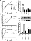
Efficient replication of the H7N9 virus in mammalian cell lines correlates with low activation of the human IFN-β promoter. (A to C) Multicyclic replication of the virus strains A/Anhui/1/2013 (hu-H7), A/Turkey-Italy/472/1999 (av-H7-It), A/Turkey-Germany/R11/2001 (av-H7-Ger), and A/Panama/2007/1999 (hu-H3) was determined in cultures of human A549 lung epithelial cells (A), in chicken DF1 fibroblasts (B), and in canine MDCK epithelial cells (C). Cells were infected with the indicated viruses (multiplicity of infection [MOI] = 0.01) and were incubated at 37°C in the presence of trypsin for 72 h. Aliquots taken at the indicated points were titrated on MDCK cells by using a standard plaque assay. The graphs show mean values and SEM from 3 independent experiments, each done with biological duplicates. Dashed lines indicate the limit of detection. Hu-H7 replicated as efficiently on A549 and MDCK cells as hu-H3, whereas av-H7 viruses were impaired in growth by about 100-fold. On DF1 cells, the viruses showed less variable replication. (D) MDCK cells with a stably integrated human IFN-β promoter luciferase reporter were mock treated or infected with hu-H7, hu-H3, or two av-H7 viruses for 12 h (MOI = 1). We used an isogenic NS1-deleted H3N2 (hu-H3 ΔNS1) mutant virus to determine reporter activation in the absence of a viral IFN antagonist (25). Cells were lysed, and equal amounts of protein were analyzed by luciferase assay (3 experiments conducted in triplicate each, mean ± SEM; top). Expression of viral NP and NS1 proteins and cellular actin as the loading control was verified by immunoblotting (bottom). Infection with hu-H3 (ΔNS1) caused a strong activation of the IFN-β promoter, whereas a modest induction was observed for all other viruses. (E) NS1 expression plasmids were constructed by placing viral NS cDNA of the indicated strains under the control of the human cytomegalovirus immediate early promoter. Human 293T cells were transfected for 24 h with an IFN-β promoter reporter plasmid, FLAG-RIG-I, and pRL-TK, expressing a renilla luciferase under the control of the constitutive herpes simplex virus (HSV) tk promoter for normalization and an NS1 expression construct from the indicated virus strain as described (25). Empty vector was used as the control. Where indicated, samples were infected for 16 h with NS1-deficient influenza virus (MOI = 1). IFN-β induction was determined by dual-luciferase assays (n = 3 in triplicates, mean ± SEM; *, P ≤ 0.05; Mann-Whitney U test). Hu-H7 NS reduced IFN-β promoter activity significantly stronger than avian H7 strains.

References

    1. Gao HN, Lu HZ, Cao B, Du B, Shang H, Gan JH, Lu SH, Yang YD, Fang Q, Shen YZ, Xi XM, Gu Q, Zhou XM, Qu HP, Yan Z, Li FM, Zhao W, Gao ZC, Wang GF, Ruan LX, Wang WH, Ye J, Cao HF, Li XW, Zhang WH, Fang XC, He J, Liang WF, Xie J, Zeng M, Wu XZ, Li J, Xia Q, Jin ZC, Chen Q, Tang C, Zhang ZY, Hou BM, Feng ZX, Sheng JF, Zhong NS, Li LJ. 2013. Clinical findings in 111 cases of influenza A (H7N9) virus infection. N. Engl. J. Med. 368:2277–2285 - PubMed
    1. WHO 12 August 2013, posting date Number of confirmed human cases of avian influenza (H7N9) reported to WHO. World Health Organization, Geneva, Switzerland: http://www.who.int/influenza/human_animal_interface/influenza_h7n9/09_Re...
    1. Chen Y, Liang W, Yang S, Wu N, Gao H, Sheng J, Yao H, Wo J, Fang Q, Cui D, Li Y, Yao X, Zhang Y, Wu H, Zheng S, Diao H, Xia S, Zhang Y, Chan KH, Tsoi HW, Teng JL, Song W, Wang P, Lau SY, Zheng M, Chan JF, To KK, Chen H, Li L, Yuen KY. 2013. Human infections with the emerging avian influenza A H7N9 virus from wet market poultry: clinical analysis and characterisation of viral genome. Lancet 381:1916–1925 - PMC - PubMed
    1. Gao R, Cao B, Hu Y, Feng Z, Wang D, Hu W, Chen J, Jie Z, Qiu H, Xu K, Xu X, Lu H, Zhu W, Gao Z, Xiang N, Shen Y, He Z, Gu Y, Zhang Z, Yang Y, Zhao X, Zhou L, Li X, Zou S, Zhang Y, Li X, Yang L, Guo J, Dong J, Li Q, Dong L, Zhu Y, Bai T, Wang S, Hao P, Yang W, Zhang Y, Han J, Yu H, Li D, Gao GF, Wu G, Wang Y, Yuan Z, Shu Y. 2013. Human infection with a novel avian-origin influenza A (H7N9) virus. N. Engl. J. Med. 368:1888–1897 - PubMed
    1. Liu D, Shi W, Shi Y, Wang D, Xiao H, Li W, Bi Y, Wu Y, Li X, Yan J, Liu W, Zhao G, Yang W, Wang Y, Ma J, Shu Y, Lei F, Gao GF. 2013. Origin and diversity of novel avian influenza A H7N9 viruses causing human infection: phylogenetic, structural, and coalescent analyses. Lancet 381:1926–1932 - PubMed

Publication types

Substances

Associated data