. 1998 Apr 28;95(9):5027-32.
doi: 10.1073/pnas.95.9.5027.

A cytoplasmic protein, bystin, interacts with trophinin, tastin, and cytokeratin and may be involved in trophinin-mediated cell adhesion between trophoblast and endometrial epithelial cells

Affiliations

A cytoplasmic protein, bystin, interacts with trophinin, tastin, and cytokeratin and may be involved in trophinin-mediated cell adhesion between trophoblast and endometrial epithelial cells

N Suzuki et al. Proc Natl Acad Sci U S A. .

Abstract

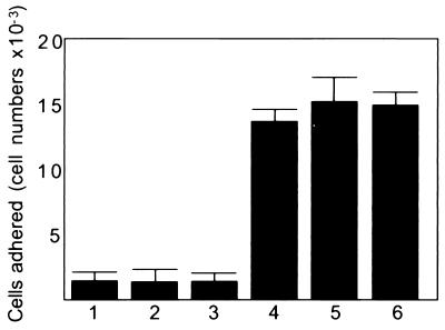
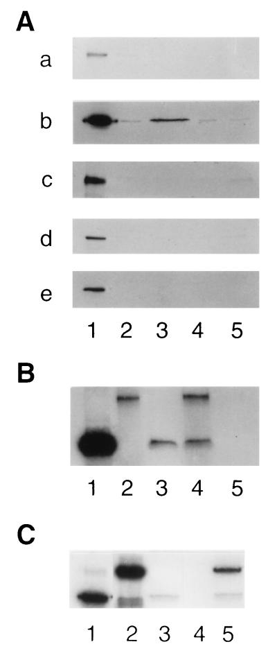
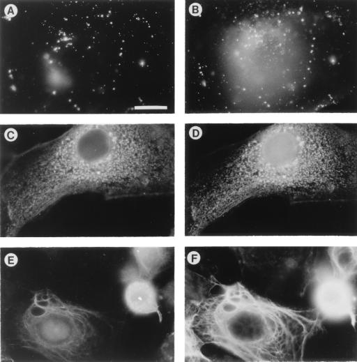
Trophinin and tastin form a cell adhesion molecule complex that potentially mediates an initial attachment of the blastocyst to uterine epithelial cells at the time of implantation. Trophinin and tastin, however, do not directly bind to each other, suggesting the presence of an intermediary protein. The present study identifies a cytoplasmic protein, named bystin, that directly binds trophinin and tastin. Bystin consists of 306 amino acid residues and is predicted to contain tyrosine, serine, and threonine residues in contexts conforming to motifs for phosphorylation by protein kinases. Database searches revealed a 53% identity of the predicted peptide sequence with the Drosophila bys (mrr) gene. Direct protein-protein interactions of trophinin, tastin, and bystin analyzed by yeast two-hybrid assays and by in vitro protein binding assays indicated that binding between bystin and trophinin and between bystin and tastin is enhanced when cytokeratin 8 and 18 are present as the third molecule. Immunocytochemistry of bystin showed that bystin colocalizes with trophinin, tastin, and cytokeratins in a human trophoblastic teratocarcinoma cell, HT-H. It is therefore possible that these molecules form a complex and thus are involved in the process of embryo implantation.


Figures

Figure 1
Figure 1
Binding of transfected COS-1 cells to the surface of monolayers of SNG-M cells. COS-1 cells were transfected with pcDNAI vector (bar 1), trophinin cDNA alone (bar 2), bystin cDNA alone (bar 3), a mixture of trophinin and bystin cDNAs (bar 4), a mixture of trophinin and tastin cDNAs (bar 5), and a mixture of trophinin, bystin and tastin cDNAs (bar 6). Two days after transfection, COS-1 cells were subjected to a cell adhesion assay. Numbers presented are the averages obtained by duplicate counting.
Figure 2
Figure 2
Comparison of human bystin and putative polypeptides homologous to bystin in D. melanogaster, C. elegans, and S. cerevisiae. (A) Alignment of amino acid residues of human bystin and the Drosophila putative bys gene product. (B) Highly homologous regions of peptide sequences among human, fly, nematode, and yeast bystins (GenBank accession nos. L02076 for D. melanogaster, U13876 for C. elegans, and Z36116 for S. cerevisiae genes).
Figure 3
Figure 3
Autoradiographs of [35S]proteins bound to GST fusion proteins in in vitro protein binding assays. GST and GST fusion proteins were produced in bacteria, immobilized on glutathione-Sepharose, and incubated with 35S-labeled in vitro-translated polypeptides produced in rabbit reticulocyte lysates. (A) [35S]Tastin (blot a), [35S]bystin (blot b), [35S]trophinin (blot c), [35S]cytokeratin 8 (blot d), and [35S]cytokeratin 18 (blot e), prepared by in vitro translation (lanes 1) were incubated with GST (lanes 2), GST-tastin (lanes 3), GST-bystin (lanes 4), or GST-trophinin (lanes 5) immobilized on glutathione-beads. After washing the beads, the bound materials were analyzed by SDS/PAGE followed by autoradiography. (B) Materials bound to GST-tastin. [35S]Bystin alone (lane 3), [35S]trophinin and [35S]bystin (lane 4), and [35S]trophinin alone (lane 5) were incubated with GST-tastin beads. Lanes 1 and 2 show [35S]bystin and [35S]trophinin used for the binding assay. (C) Materials bound to GST-tastin. [35S]Bystin alone (lane 3), [35S]cytokeratin 18 alone (lane 4), and [35S]cytokeratin 18 and [35S]bystin (lane 5) were incubated with GST-tastin beads. Lanes 1 and 2 show [35S]bystin and [35S]cytokeratin 18 used for the binding assay.
Figure 4
Figure 4
Immunofluorescence micrographs of HT-H cells stained with antibodies for trophinin, bystin, tastin, and cytokeratin. (A and B) Double immunostaining showing trophinin (A) and bystin (B). Unpermeabilized cells were stained with fluorescein isothiocyanate (FITC)-conjugated anti-trophinin antibody (A), permeabilized, and stained with rhodamine-conjugated anti-bystin antibody (B). These photographs focused on the upper cell surface where trophinin is present. Accordingly, only a fraction of bystin localized near upper plasma membranes is shown. (C and D) Double immunostaining showing bystin (C) and tastin (D). Cells were permeabilized and stained with unconjugated anti-bystin and anti-tastin antibodies, followed by FITC-conjugated goat anti-mouse IgM antibody for bystin (C) and rhodamine-conjugated goat anti-mouse IgG antibody for tastin (D). (E and F) Double immunostaining showing bystin (E) and cytokeratin 8 (F). Cells were permeabilized and stained with unconjugated anti-bystin and anti-cytokeratin antibodies, followed by FITC-conjugated goat anti-mouse IgM antibody for bystin (E) and rhodamine conjugated goat anti-rat IgG antibody for cytokeratin 8 (F). All photographs are presented at the same magnification. (Bar = 20 μm.)

References

    1. Schlafke S, Enders A C. Biol Reprod. 1975;12:41–65. - PubMed
    1. Denker H W. In: Trophoblast Research. Denker H W, Aplin J D, editors. New York: Plenum; 1990. pp. 3–20.
    1. Psychoyos A. In: Handbook of Physiology. Greep R O, Astwood E G, Geige R G, editors. Washington, DC: Am. Physiol. Soc.; 1973. pp. 187–215.
    1. Day S K. In: Reproductive Endocrinology, Surgery and Technology. Adashi E Y, Rock J A, Rosenwaks Z, editors. Philadelphia: Lippincott–Raven; 1996. pp. 421–434.
    1. Paria B C, Huet-Hudson Y M, Dey S K. Proc Natl Acad Sci USA. 1993;90:10159–10162. - PMC - PubMed

Publication types

Associated data