. 2014 Nov 20:5:5560.
doi: 10.1038/ncomms6560.

Microhomology-mediated end-joining-dependent integration of donor DNA in cells and animals using TALENs and CRISPR/Cas9

Affiliations

Microhomology-mediated end-joining-dependent integration of donor DNA in cells and animals using TALENs and CRISPR/Cas9

Shota Nakade et al. Nat Commun. .

Abstract

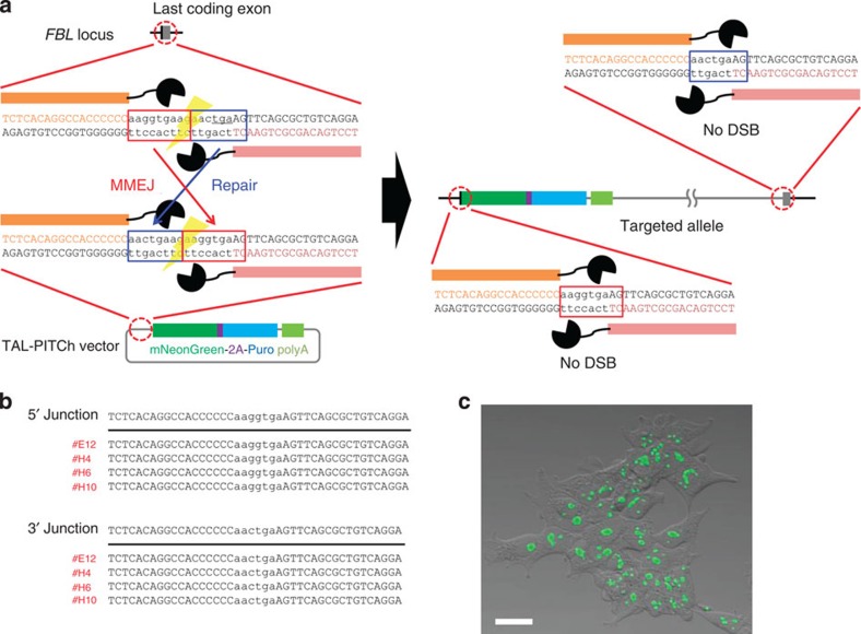
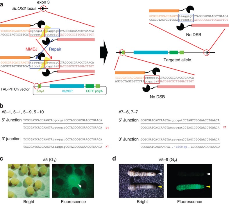
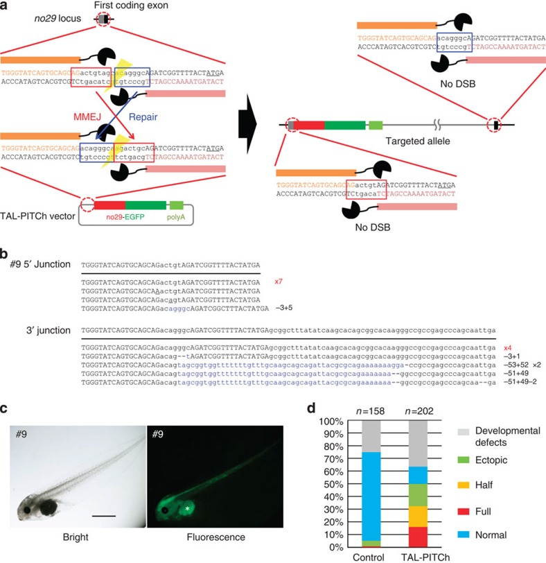
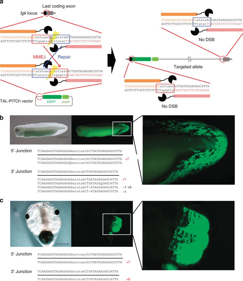
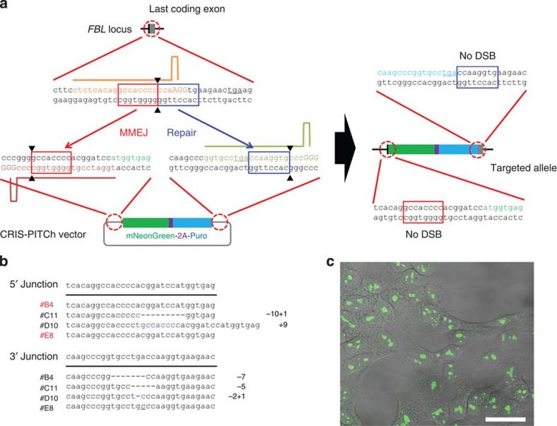
Genome engineering using programmable nucleases enables homologous recombination (HR)-mediated gene knock-in. However, the labour used to construct targeting vectors containing homology arms and difficulties in inducing HR in some cell type and organisms represent technical hurdles for the application of HR-mediated knock-in technology. Here, we introduce an alternative strategy for gene knock-in using transcription activator-like effector nucleases (TALENs) and clustered regularly interspaced short palindromic repeats (CRISPR)/CRISPR-associated 9 (Cas9) mediated by microhomology-mediated end-joining, termed the PITCh (Precise Integration into Target Chromosome) system. TALEN-mediated PITCh, termed TAL-PITCh, enables efficient integration of exogenous donor DNA in human cells and animals, including silkworms and frogs. We further demonstrate that CRISPR/Cas9-mediated PITCh, termed CRIS-PITCh, can be applied in human cells without carrying the plasmid backbone sequence. Thus, our PITCh-ing strategies will be useful for a variety of applications, not only in cultured cells, but also in various organisms, including invertebrates and vertebrates.


Figures

Figure 1
Figure 1. TAL-PITCh in human cells.
(a) Schematic illustration of TAL-PITCh at the human FBL locus. Orange and pink letters indicate the left and right TALEN target sites, respectively. Red and blue boxes indicate the microhomologous sequences. The stop codon is underlined. (b) Sequences of knocked-in clones. The intended knocked-in sequence is shown at the top. TALEN target sites are shown in capital letters. Red letters indicate correctly knocked-in clones. (c) Confocal laser scanning microscopy image of knocked-in cells showing nucleolar localization of mNeonGreen fluorescence (clone #H6). Scale bar, 30 μm.
Figure 2
Figure 2. TAL-PITCh in silkworms.
(a) Schematic illustration of TAL-PITCh at the B. mori BLOS2 locus. Orange and pink letters indicate the left and right TALEN target sites, respectively. Red and blue boxes indicate the microhomologous sequences. hsp90P, hsp90 promoter. (b) Sequences of knocked-in alleles from the six G1 worms (#2–1, #5–1, #5–9, #5–10, #7–6 and #7–7). The intended knocked-in sequence is shown at the top. TALEN target sites are shown in capital letters. Red letters indicate correctly knocked-in alleles. Blue letters indicate insertions. (c) Bright-field and fluorescence microscopy images of the G1 embryos in the #5 batch. Strong EGFP expression could be observed in the putative knock-in embryo, according to the activity of the hsp90 promoter (arrowhead). Scale bar, 0.5 mm. (d) Bright-field and fluorescence microscopy images of the G2 larva derived from #5–9 batch. White and yellow arrowheads indicate wild-type and knock-in silkworms, respectively. Note that the knock-in larva shows an oily skin phenotype. Scale bar, 1 mm.
Figure 3
Figure 3. TAL-PITCh at the no29 locus in frog embryos.
(a) Schematic illustration of TAL-PITCh at the X. laevis no29 locus. Orange and pink letters indicate the left and right TALEN target sites, respectively. Red and blue boxes indicate the microhomologous sequences. The start codons are underlined. (b) Sequences of knocked-in alleles from embryo #9. The intended knocked-in sequence is shown at the top. TALEN target sites are shown in capital letters. Red letters indicate correctly knocked-in alleles. Blue letters indicate insertions. Dashes indicate deletions. Substitutions are underlined. (c) Bright-field and fluorescence microscopy images of embryo #9. An asterisk indicates yolk autofluorescence. Scale bar, 1 mm. (d) Percentage of phenotypes in the control embryos and the TAL-PITChed embryos. For the control, the vector from which the TALEN target site was removed was used instead of the TAL-PITCh vector. Except for abnormally developed embryos, phenotypes were divided into four groups (full, half, ectopic and normal) according to the expressed region of EGFP. Total numbers of individuals are shown at the top of each graph.
Figure 4
Figure 4. TAL-PITCh at the fgk locus in frog embryos.
(a) Schematic illustration of TAL-PITCh at the X. laevis fgk locus. Orange and pink letters indicate the left and right TALEN target sites, respectively. Red and blue boxes indicate the microhomologous sequences. The stop codon is underlined. (b,c) Microscopic images and sequences of the TAL-PITChed embryos showing EGFP expression in the fin (b) and the gill (c). TALEN target sites are shown in capital letters. Red letters indicate correctly knocked-in alleles. Substitutions are underlined. Scale bars, 1 mm.
Figure 5
Figure 5. CRIS-PITCh in human cells.
(a) Schematic illustration of CRISPR/Cas9-mediated targeted integration using CRIS-PITCh. Orange, pink and green letters indicate the gRNA target sites. Red and blue boxes indicate the microhomologous sequences. The stop codons are underlined. (b) Sequences of knocked-in clones. The intended knocked-in sequence is shown at the top. Dashes indicate deletions. Blue letters indicate the insertion. The substitution is underlined. (c) Confocal laser scanning microscopy image of knocked-in cells showing nucleolar localization of mNeonGreen fluorescence (clone #B4). Scale bar, 30 μm.

References

    1. Carroll D. Genome engineering with targetable nucleases. Annu. Rev. Biochem. 83, 409–439 (2014). - PubMed
    1. Kim H. & Kim J. S. A guide to genome engineering with programmable nucleases. Nat. Rev. Genet. 15, 321–334 (2014). - PubMed
    1. Chen F. et al. High-frequency genome editing using ssDNA oligonucleotides with zinc-finger nucleases. Nat. Methods 8, 753–755 (2011). - PMC - PubMed
    1. Miyaoka Y. et al. Isolation of single-base genome-edited human iPS cells without antibiotic selection. Nat. Methods 11, 291–293 (2014). - PMC - PubMed
    1. Hockemeyer D. et al. Efficient targeting of expressed and silent genes in human ESCs and iPSCs using zinc-finger nucleases. Nat. Biotechnol. 27, 851–857 (2009). - PMC - PubMed

Publication types