. 2020 Jun;287(12):2612-2630.
doi: 10.1111/febs.15154. Epub 2019 Dec 24.

A GM1b/asialo-GM1 oligosaccharide-binding R-type lectin from purplish bifurcate mussels Mytilisepta virgata and its effect on MAP kinases

Affiliations

A GM1b/asialo-GM1 oligosaccharide-binding R-type lectin from purplish bifurcate mussels Mytilisepta virgata and its effect on MAP kinases

Yuki Fujii et al. FEBS J. 2020 Jun.

Abstract

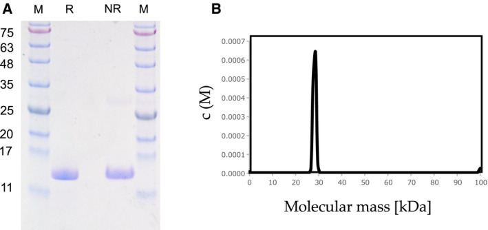
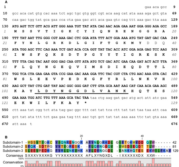
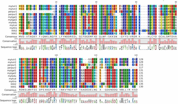
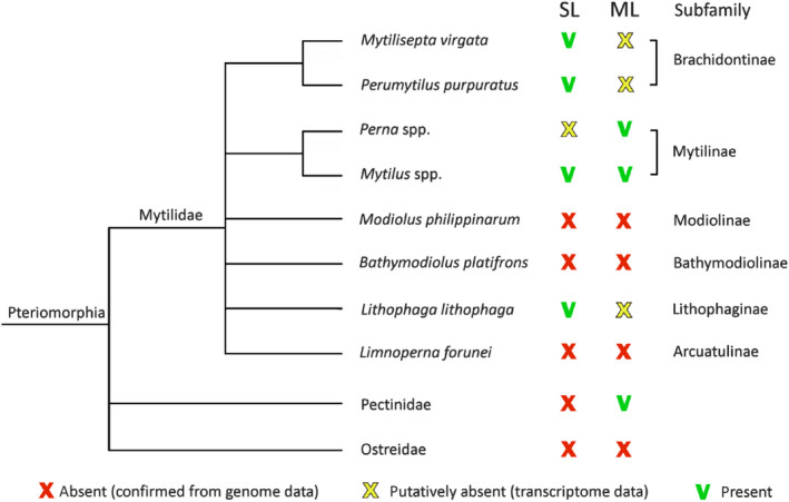
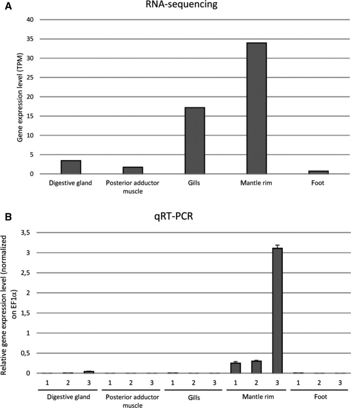
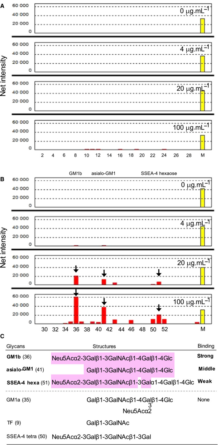
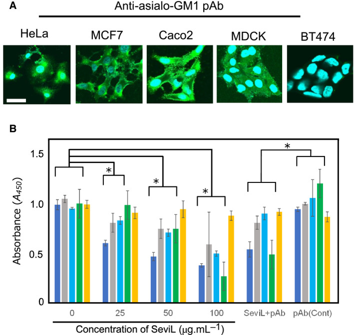
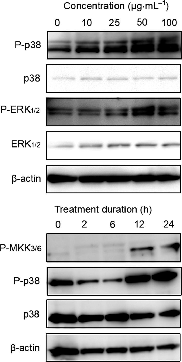
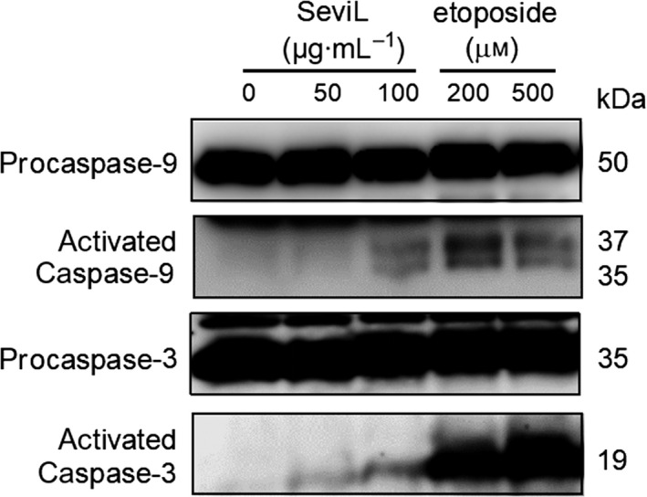
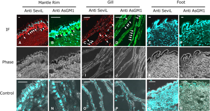
A 15-kDa lectin, termed SeviL, was isolated from Mytilisepta virgata (purplish bifurcate mussel). SeviL forms a noncovalent dimer that binds strongly to ganglio-series GM1b oligosaccharide (Neu5Acɑ2-3Galβ1-3GalNAcβ1-4Galβ1-4Glc) and its precursor, asialo-GM1 (Galβ1-3GalNAcβ1-4Galβ1-4Glc). SeviL also interacts weakly with the glycan moiety of SSEA-4 hexaose (Neu5Acα2-3Galβ1-3GalNAcβ1-3Galα1-4Galβ1-4Glc). A partial protein sequence of the lectin was determined by mass spectrometry, and the complete sequence was identified from transcriptomic analysis. SeviL, consisting of 129 amino acids, was classified as an R(icin B)-type lectin, based on the presence of the QxW motif characteristic of this fold. SeviL mRNA is highly expressed in gills and, in particular, mantle rim tissues. Orthologue sequences were identified in other species of the family Mytilidae, including Mytilus galloprovincialis, from which lectin MytiLec-1 was isolated and characterized in our previous studies. Thus, mytilid species contain lectins belonging to at least two distinct families (R-type lectins and mytilectins) that have a common β-trefoil fold structure but differing glycan-binding specificities. SeviL displayed notable cytotoxic (apoptotic) effects against various cultured cell lines (human breast, ovarian, and colonic cancer; dog kidney) that possess asialo-GM1 oligosaccharide at the cell surface. This cytotoxic effect was inhibited by the presence of anti-asialo-GM1 oligosaccharide antibodies. With HeLa ovarian cancer cells, SeviL showed dose- and time-dependent activation of kinase MKK3/6, p38 MAPK, and caspase-3/9. The transduction pathways activated by SeviL via the glycosphingolipid oligosaccharide were triggered apoptosis. DATABASE: Nucleotide sequence data have been deposited in the GenBank database under accession numbers MK434191, MK434192, MK434193, MK434194, MK434195, MK434196, MK434197, MK434198, MK434199, MK434200, and MK434201.

Keywords: Mytilisepta virgata; R-type lectin; bivalves; ganglioside; purplish bifurcate mussels.


Conflict of interest statement

The authors declare no conflict of interest.

Figures

Figure 1
Figure 1
Purification of Mytilisepta virgata lectin, SeviL. (A) SDS/PAGE pattern under reducing (R) and nonreducing (NR) conditions. Numbers on the left indicate molecular masses (kDa) of marker proteins (M). (B) The molecular weight of the native protein (30 kDa) obtained from distribution of sedimentation coefficient by sedimentation velocity AUC. It indicates the presence of dimers in solution, with negligible amounts of monomer. Concentration c(M) was measured in absorption units (A280).
Figure 2
Figure 2
cDNA sequence and deduced amino acid sequence of SeviL. (A) The asterisk indicates the stop codon. The peptide fragment obtained from mass spectrometric analysis is underlined. (B) Amino acid sequence alignment of the internal tandem‐repeat subdomains of SeviL. The consensus at bottom summarizes the residues shared by the three domains. The sequence alignment within the polypeptide is analyzed by using MUSCLE program 31.
Figure 3
Figure 3
Multiple sequence alignment of SeviL orthologues in various mytilid species, detected in publicly available genomic or transcriptomic resources. mytvir1 (GenBank accession number MK434191) and mytvir2 (MK434192): Mytilisepta virgata; mytgal1 (MK434193) and mtgel2 (MK434194): Mytilus galloprovincialis; litlit1 (MK434195) and litlit2 (MK434196): Lithophaga lithophaga; mytedu (MK434197): Mytilus edulis; mytcal (MK434198): Mytilus californianus; myttro (MK434199): Mytilus trossulus; perpur1 (MK434200) and perpur2 (MK434201): Perumytilus purpuratus. Names with numbers (e.g., mytvir1, mytvir2) indicate lectin variants from the same organism. Note that the sequence perpur1 and perpur2 display incomplete N‐terminal and C‐terminal regions, respectively. The sequence alignment within the polypeptide is analyzed by using MUSCLE program 31.
Figure 4
Figure 4
Comparative distribution of SeviL/R‐type (SL) lectin and mytilectin (ML) family members in bivalves. The presence of ML and SL was confirmed by genomic and/or transcriptomic data in the family Mytilidae, using Pectinidae, and Ostreidae as outgroups in the subclass Pteriomorphia. The cladogram was drawn based on the classification of bivalve species based on WoRMS data 33ription levels of SeviL in various M. virgata tissues, calculated in silico from RNA‐seq data in a pool of individuals and expressed as trascripts per milion (TPM) (panel A), or determined in single individuals with qRT/PCR (panel B) (see Materials and methods). Each bar represents the mean plus standard deviation of three technical replicates.
Figure 5
Figure 5
Transcription levels of SeviL in various M. virgata tissues, calculated in silico from RNA‐seq data in a pool of individuals and expressed as trascripts per milion (TPM) (panel A), or determined in single individuals with qRT/PCR (panel B) (see Materials and methods). Each bar represents the mean plus standard deviation of three technical replicates.
Figure 6
Figure 6
Glycan‐binding profile of SeviL. HyLite555‐labeled SeviL (0–100 μg·mL−1: right upper) was subjected to glycan array analysis combining glycan‐conjugated chips in which 52 glycan structures were immobilized and a surface plasmon resonance scanning detector (numbering as in Fig. S3A and Table S1). The evanescent‐field fluorescence occurring by the binding between HyLite555‐SeviL and the glycans (Fig. S3B) is represented as net intensities (y‐axis of these graphs). (A) The chip includes N‐glycans, O‐glycans, glycosaminoglycans, Lewis type oligosaccharides, derivatives of lactose and N‐acetyllactosamine and ABH‐type oligosaccharides (No. 1‐28 in Table S1). (B) The chip includes ganglio‐series oligosaccharides and globo‐series oligosaccharides (No. 29‐52 in Table S1). Arrows indicate positions (glycans number 36, 41 and 51) of glycans of GM1b, asialo‐GM1, and SSEA‐4 hexsaose, respectively. M. Internal standard marker for fluorescence. C. The structural motif of the glycans recognized by SeviL. The colored part highlights the structure shared by GM1b, asialo‐GM1 and SSEA‐4 hexa(ose), whose glycans are bound by SeviL (see panel B). GM1a, TF, and SSEA‐4 tetra(ose) are glycans that are not bound by the lectin.
Figure 7
Figure 7
Detection of asialo‐GM1 expression on cell membranes by immunostaining. (A) Paraformaldehyde‐fixed cells were stained with anti‐asialo‐GM1 oligosaccharide pAb and AF488‐tagged goat anti‐rabbit Ab (green). Nuclei were counterstained with DAPI (blue). Magnification: ×40. Scale bar: 10 mm. (B) Cytotoxic effects of SeviL on human ovarian cancer (HeLa), breast cancer (MCF7, BT474), colonic cancer (Caco2), and dog kidney (MDCK) cells. Cells were treated with SeviL at various concentrations (0–100 μg·mL−1) for 24 h, and cell viability (expressed as A450; see M & M/ ‘Cell viability and cytotoxicity assays’) was determined by WST‐8 assay. SeviL + pAb: treatment with 50 μg·mL−1 SeviL plus anti‐asialo‐GM1 oligosaccharide polyclonal antibody (pAb). pAb (Cont): anti‐asialo‐GM1 oligosaccharide pAb without SeviL, as control. Data shown are mean ± SE (n = 3). P values (*P < 0.05) were obtained with Dunnett’s test.
Figure 8
Figure 8
Phosphorylation of MAPKs by SeviL in HeLa cells. Cells (5 × 105) were treated with SeviL at various concentration (0–100 μg·mL−1) or durations (0–24 h). Phosphorylation of kinases was evaluated by Western blotting of cell lysates. P‐p38, P‐ERK1/2, and P‐MKK3/6: phosphorylated forms of p38, ERK1/2 and MKK3/6 kinase, respectively. MKK3/6 is the upstream kinase of ERK1/2.
Figure 9
Figure 9
Induction of caspase‐3/9 cleavage by SeviL. HeLa cells were treated for 48 h with SeviL (50, 100 μg·mL−1), anticancer reagent etoposide (200, 500 μm) or lysates were prepared, and caspase‐3/9 activation was detected by Western blotting. Each experiment was performed in triplicate.
Figure 10
Figure 10
Localization of SeviL and substances which reacted with anti‐asialo‐GM1 polyclonal antibody in M. virgata tissues. Tissue sections derived from mantle rim, gill, and foot were applied by anti‐SeviL (column anti‐SeviL: A, C, E, G, I and K) and anti‐asialo‐GM1 (column Anti AsGM1: B, D, F, H, J, and I) polyclonal antibodies followed by Alexa 568 (red: for the detection Anti‐SeviL) or Alexa488 (green: for the detection of As asialo‐GM1) conjugated secondary anti‐rabbit IgG antibody. All sections were stained also with DAPI to detect nuclei (blule: A–F and M‐R). IF: immunofluoresence (A–F), phase: phase contrast (G–L) and control: applying nonimmune primary antibody (for SeviL: M, O, and Q) and without primary antibody (for asialo‐GM1 antibody reactant: N, P and R), respectively. Scale bars: 20 μm each.

Similar articles

Cited by

References

    1. Ogawa T, Watanabe M, Naganuma T & Muramoto K (2011) Diversified carbohydrate‐binding lectins from marine resources. J Amino Acids 2011, 1–20. - PMC - PubMed
    1. Cheung RC, Wong JH, Pan W, Chan YS, Yin C, Dan X & Ng TB (2015) Marine lectins and their medicinal applications. Appl Microbiol Biotechnol 99, 3755–3773. - PMC - PubMed
    1. Chernikov OV, Molchanova VI, Chikalovets IV, Kondrashina AS, Li W & Lukyanov PA (2013) Lectins of marine hydrobionts. Biochemistry (Mosc) 78, 760–770. - PMC - PubMed
    1. Rao A, Seto J, Berg JK, Kreft SG, Scheffner M & Cölfen H (2013) Roles of larval sea urchin spicule SM50 domains in organic matrix self‐assembly and calcium carbonate mineralization. J Struct Biol 183, 205–215. - PubMed
    1. Motohashi S, Jimbo M, Naito T, Suzuki T, Sakai R & Kamiya H (2017) Isolation, amino acid sequences, and plausible functions of the galacturonic acid‐binding egg lectin of the sea hare Aplysia kurodai . Mar Drugs 15, 161. - PMC - PubMed

Publication types