Exon skipping and dystrophin restoration in patients with Duchenne muscular dystrophy after systemic phosphorodiamidate morpholino oligomer treatment: an open-label, phase 2, dose-escalation study
- PMID: 21784508
- PMCID: PMC3156980
- DOI: 10.1016/S0140-6736(11)60756-3
Exon skipping and dystrophin restoration in patients with Duchenne muscular dystrophy after systemic phosphorodiamidate morpholino oligomer treatment: an open-label, phase 2, dose-escalation study
Abstract
Background: We report clinical safety and biochemical efficacy from a dose-ranging study of intravenously administered AVI-4658 phosphorodiamidate morpholino oligomer (PMO) in patients with Duchenne muscular dystrophy.
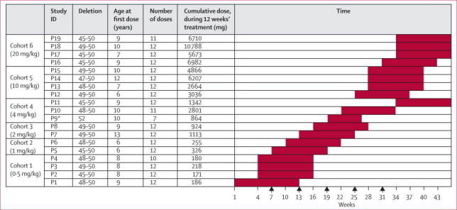
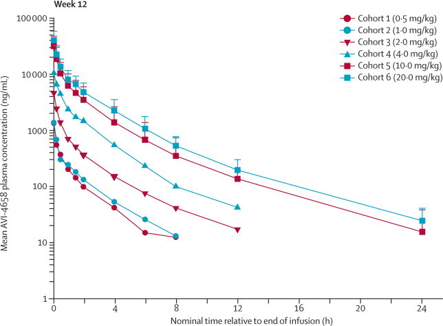
Method: We undertook an open-label, phase 2, dose-escalation study (0·5, 1·0, 2·0, 4·0, 10·0, and 20·0 mg/kg bodyweight) in ambulant patients with Duchenne muscular dystrophy aged 5-15 years with amenable deletions in DMD. Participants had a muscle biopsy before starting treatment and after 12 weekly intravenous infusions of AVI-4658. The primary study objective was to assess safety and tolerability of AVI-4658. The secondary objectives were pharmacokinetic properties and the ability of AVI-4658 to induce exon 51 skipping and dystrophin restoration by RT-PCR, immunohistochemistry, and immunoblotting. The study is registered, number NCT00844597.
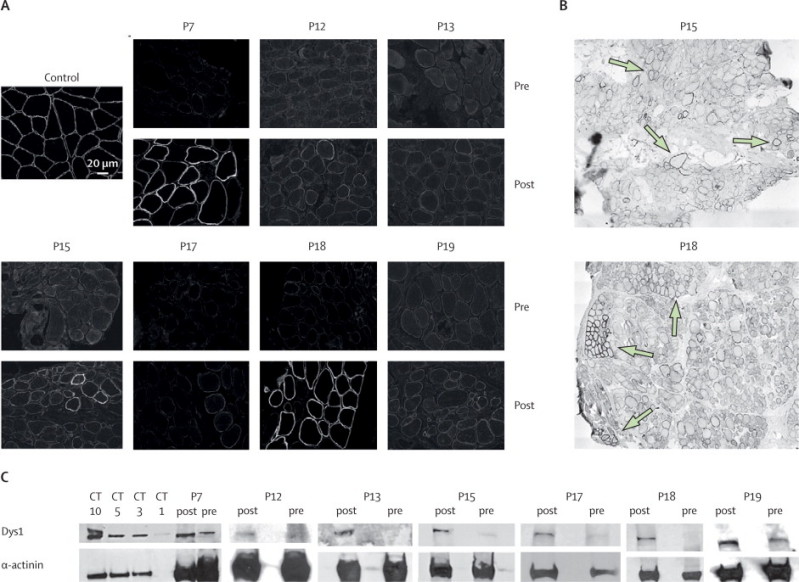
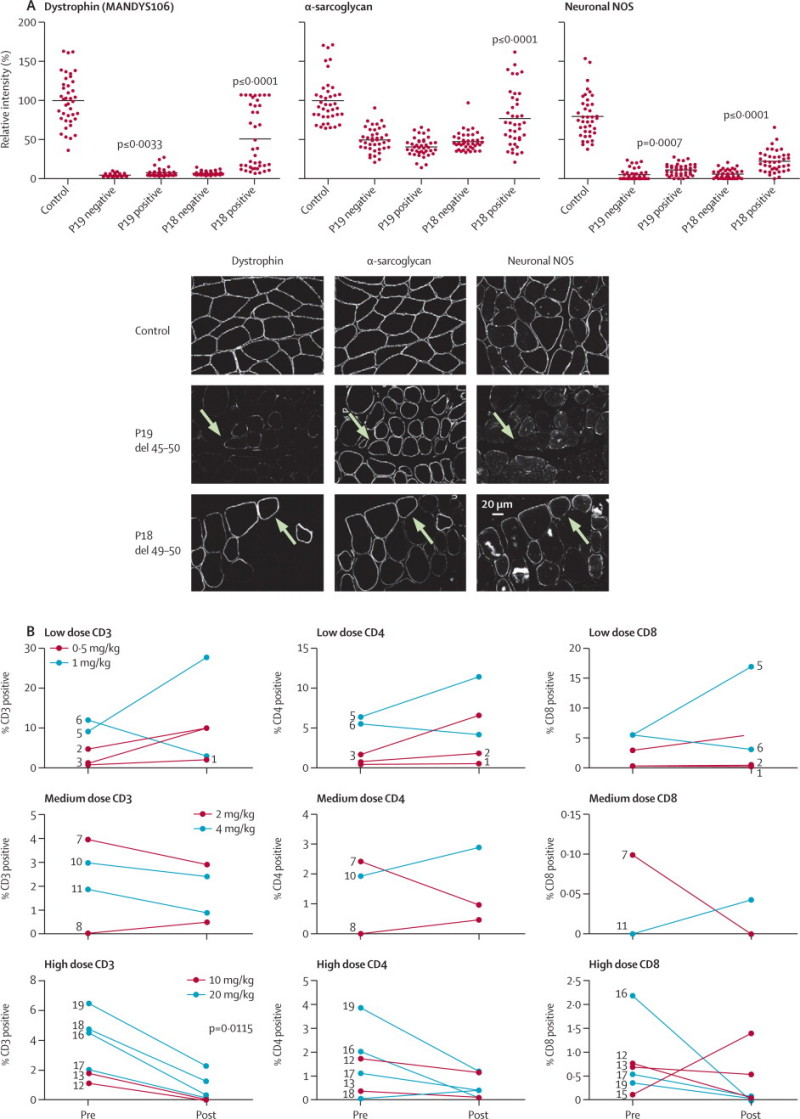
Findings: 19 patients took part in the study. AVI-4658 was well tolerated with no drug-related serious adverse events. AVI-4658 induced exon 51 skipping in all cohorts and new dystrophin protein expression in a significant dose-dependent (p=0·0203), but variable, manner in boys from cohort 3 (dose 2 mg/kg) onwards. Seven patients responded to treatment, in whom mean dystrophin fluorescence intensity increased from 8·9% (95% CI 7·1-10·6) to 16·4% (10·8-22·0) of normal control after treatment (p=0·0287). The three patients with the greatest responses to treatment had 21%, 15%, and 55% dystrophin-positive fibres after treatment and these findings were confirmed with western blot, which showed an increase after treatment of protein levels from 2% to 18%, from 0·9% to 17%, and from 0% to 7·7% of normal muscle, respectively. The dystrophin-associated proteins α-sarcoglycan and neuronal nitric oxide synthase were also restored at the sarcolemma. Analysis of the inflammatory infiltrate indicated a reduction of cytotoxic T cells in the post-treatment muscle biopsies in the two high-dose cohorts.
Interpretation: The safety and biochemical efficacy that we present show the potential of AVI-4658 to become a disease-modifying drug for Duchenne muscular dystrophy.
Funding: UK Medical Research Council; AVI BioPharma.
Copyright © 2011 Elsevier Ltd. All rights reserved.
Figures
Comment in
-
Exon-skipping therapy for Duchenne muscular dystrophy.Lancet. 2011 Aug 13;378(9791):546-7. doi: 10.1016/S0140-6736(11)61028-3. Epub 2011 Jul 23. Lancet. 2011. PMID: 21784507 No abstract available.
-
Skipping along: an exon skipping therapy shows promise for Duchenne muscular dystrophy.Clin Genet. 2011 Nov;80(5):424-5. doi: 10.1111/j.1399-0004.2011.01769.x. Epub 2011 Sep 12. Clin Genet. 2011. PMID: 21883166 No abstract available.
-
Exon-skipping therapy for Duchenne muscular dystrophy.Lancet. 2012 Jan 14;379(9811):e10; author reply e10-11. doi: 10.1016/S0140-6736(12)60063-4. Lancet. 2012. PMID: 22243826 No abstract available.
References
-
- Bushby K, Finkel R, Birnkrant DJ, for the DMD Care Considerations Working Group Diagnosis and management of Duchenne muscular dystrophy, part 1: diagnosis, and pharmacological and psychosocial management. Lancet Neurol. 2010;9:77–93. - PubMed
-
- Hoffman EP, Brown RH, Jr, Kunkel LM. Dystrophin: the protein product of the Duchenne muscular dystrophy locus. Cell. 1987;51:919–928. - PubMed
-
- Bushby KM, Gardner-Medwin D, Nicholson LV. The clinical, genetic and dystrophin characteristics of Becker muscular dystrophy. II. Correlation of phenotype with genetic and protein abnormalities. J Neurol. 1993;240:105–112. - PubMed
-
- Sazani P, Graziewicz MA, Kole R. Splice switching oligonucleotides as potential therapeutics. In: Crooke ST, editor. Antisense drug technology, principles, strategies and applications. CBC Press; Boca Raton, FL, USA: 2008. pp. 89–114.
Publication types
MeSH terms
Substances
Associated data
Grants and funding
LinkOut - more resources
Full Text Sources
Other Literature Sources
Medical