

F18-choline PET/CT guided surgery in primary hyperparathyroidism when ultrasound and MIBI SPECT/CT are negative or inconclusive: the APACH1 study
- PMID: 29270788
- PMCID: PMC5829113
- DOI: 10.1007/s00259-017-3911-1
F18-choline PET/CT guided surgery in primary hyperparathyroidism when ultrasound and MIBI SPECT/CT are negative or inconclusive: the APACH1 study
Abstract
Purpose: To evaluate the sensitivity of F18-choline (FCH) PET/CT for parathyroid adenoma detection prior to surgery in patients with primary hyperparathyroidism and negative or inconclusive cervical ultrasound and Tc99m-sestaMIBI SPECT/CT.
Methods: We conducted a prospective bicentric study (NCT02432599). All patients underwent FCH PET/CT. The result was scored positive, inconclusive or negative. The number of uptakes and their sites were recorded. The FCH PET/CT result guided the surgical procedure (minimally invasive parathyroidectomy, bilateral cervical exploration, or other in case of multiple or ectopic foci). FCH PET/CT results were compared to the surgical and pathological findings and the follow-up.
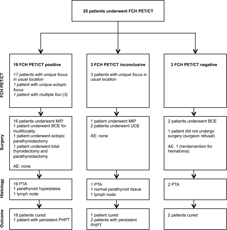
Results: Twenty-five patients were included. Mean calcium and PTH levels prior to surgery were 2.76 ± 0.17 mmol/l and 94.8 ± 37.4 ng/l. Nineteen (76%) FCH PET/CTs were scored positive, 3 (12%) inconclusive and 3 (12%) negative, showing 21 cases of uniglandular disease, including 1 ectopic localization and 1 case of multiglandular (3 foci) disease. Mean lesion size was 13.1 ± 8.6 mm. Twenty-four patients underwent surgery. FCH PET/CT guided surgery in 22 (88%) patients, allowing for 17 minimally invasive parathyroidectomies, 1 bilateral cervical exploration for multifocality and 4 other surgical procedures. Two patients with negative FCH-PET/CT underwent bilateral cervical exploration. When dichotomizing the FCH PET/CT results, thereby classifying the inconclusive FCH PET/CT results as positive, the per lesion and per patient sensitivities were 91.3% (95%CI: 72.0-98.9) and 90.5% (95%CI: 69.6-98.8) and the corresponding positive predictive values were 87.5% (95%CI: 67.6-97.3) and 86.4% (95%CI: 65.1-97.1), respectively. Twenty-one (88%) patients were considered cured after surgery. Their mean calcium level after surgery was 2.36 ± 0.17 mmol/l.
Conclusions: Preoperative FCH PET/CT has a high sensitivity and positive predictive value for parathyroid adenoma detection in patients with primary hyperparathyroidism and negative or inconclusive conventional imaging results. Bilateral cervical exploration could be avoided in the majority (75%) of patients.
Keywords: F18-choline; MIBI SPECT/CT; Minimally invasive surgery; PET/CT; Parathyroid adenoma; Primary hyperparathyroidism.
Conflict of interest statement
Conflict of interest
The authors declare that they have no conflict of interest.
Ethical approval
All procedures performed in studies involving human participants were in accordance with the ethical standards of the institutional and/or national research committee and with the 1964 Helsinki declaration and its later amendments or comparable ethical standards.
Informed consent
Informed consent was obtained from all individual participants included in the study (Ref. 2014–41, Comité de protection des personnes Nord-Ouest III).
Figures
Comment in
-
18F-Fluorocholine PET/CT as a second line nuclear imaging technique before surgery for primary hyperparathyroidism.Eur J Nucl Med Mol Imaging. 2018 Apr;45(4):654-657. doi: 10.1007/s00259-017-3920-0. Eur J Nucl Med Mol Imaging. 2018. PMID: 29335763 No abstract available.
References
-
- Alexandrides TK, Kouloubi K, Vagenakis AG, Yarmenitis S, Spyridonidis T, Vassilakos P, et al. The value of scintigraphy and ultrasonography in the preoperative localization of parathyroid glands in patients with primary hyperparathyroidism and concomitant thyroid disease. Hormones (Athens ) 2006;5:42–51. doi: 10.14310/horm.2002.11167. - DOI - PubMed
-
- Bhansali A, Masoodi SR, Bhadada S, Mittal BR, Behra A, Singh P. Ultrasonography in detection of single and multiple abnormal parathyroid glands in primary hyperparathyroidism: comparison with radionuclide scintigraphy and surgery. Clin Endocrinol. 2006;65:340–345. doi: 10.1111/j.1365-2265.2006.02601.x. - DOI - PubMed
-
- Harari A, Mitmaker E, Grogan RH, Lee J, Shen W, Gosnell J, et al. Primary hyperparathyroidism patients with positive preoperative sestamibi scan and negative ultrasound are more likely to have posteriorly located upper gland adenomas (PLUGs) Ann Surg Oncol. 2011;18:1717–1722. doi: 10.1245/s10434-010-1493-2. - DOI - PMC - PubMed
MeSH terms
Substances
Associated data
LinkOut - more resources
Full Text Sources
Other Literature Sources
Medical
Research Materials
Miscellaneous
